Case Report

신이식 환자에서 자발적으로 발생한 저구연산뇨증 연관성 요관 결석로

나정구, 공성민, 이동규, 김성민, 장윤정, 김성록, 이유지
Author Information & Copyright
성균관대학교 의과대학 삼성창원병원 내과
Department of Internal Medicine, Samsung Changwon Hospital, Sungkyunkwan University School of Medicine, Changwon, Korea
Corresponding author Yu-Ji Lee Department of Internal Medicine, Samsung Changwon Hospital, Sungkyunkwan University School of Medicine, 158 Paryong-ro, Masanhoewon-gu, Changwon 51353, Korea Tel: 82-55-290-6332, Fax: 82-55-290-1064 E-mail: yuji.lee@samsung.com

Copyright © 2015 Ewha Womans University School of Medicine. This is an open-access article distributed under the terms of the Creative Commons Attribution Non-Commercial License (http://creativecommons.org/licenses/by-nc/4.0) which permits unrestricted non-commercial use, distribution, and reproduction in any medium, provided the original work is properly cited.

Received: May 16, 2015; Accepted: Aug 20, 2015

Published Online: Oct 31, 2015

Abstract

Urolithiasis is an uncommon complication in renal transplantation. We report a case of hypocitraturia-related ureteral steinstrasse which was spontaneously formed in a renal transplant recipient. The patient who underwent renal transplantation was admitted with acute pyelonephritis. Hydronephrosis in the transplanted kidney and multiple stones (steinstrasse) in the distal ureter were incidentally found on computed tomography scanning. After a failed attempt of ureteroscopic removal of stones, the patient underwent open ureterolithotomy and ureteroureterostomy. On stone analysis, carbonate apatite was confirmed. Urinary citric acid levels were decreased to 127.6 mg/day. Potassium citrate was administered to prevent stone recurrence by increasing urinary citrate excretion. No recurrence of stones was shown six months later. Urolithiasis in renal transplant recipients requires a high index of suspicion. Hypocitraturia can increase the risk for urolithiasis. Rapid recognition by careful surveillance, prompt removal of stones, and precautionary efforts to prevent recurrence are needed.

Keywords: Citric acid; Renal transplantation; Ureter; Stones

서 론

신이식 후 요로결석의 발생 빈도는 대략 1–2%로 비교적 드문 합병증 중 하나이다[1-3]. 이식 후 요로결석이 요로감염이나 요로폐쇄를 일으킬 수 있고, 이식 신 기능부전의 위험을 증가시킬 수 있다[2,4]. 그렇지만 신이식 환자에서 요로결석의 임상 양상이 비신이식 환자와 차이가 있는데 이식신 및 이식 뇨관의 신경차단으로 전형적인 요로결석의 통증이 없어 진단이 늦어질 수 있다[1,3]. 요관 결석로(steinstrasse)는 여러 개의 결석이 요관에 동시에 존재하는 것으로 전형적으로 체외충격파쇄석술의 합병증으로 발생하며, 자연적으로 발생하는 경우는 드물다[5]. 더욱이 신이식 환자에서 자연적으로 발생한 요관 결석로에 대해 보고된 바가 없다. 최근 저자들은 신이식 후 저구연산뇨증과 관련된 요관 결석로가 발생하여 개방 수술과 경구 용해제로 치료한 1예를 경험하였기에 보고하는 바이다.

증 례

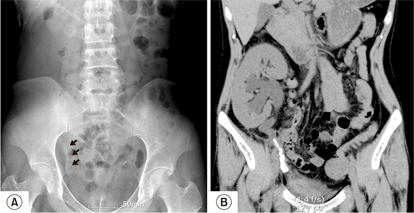
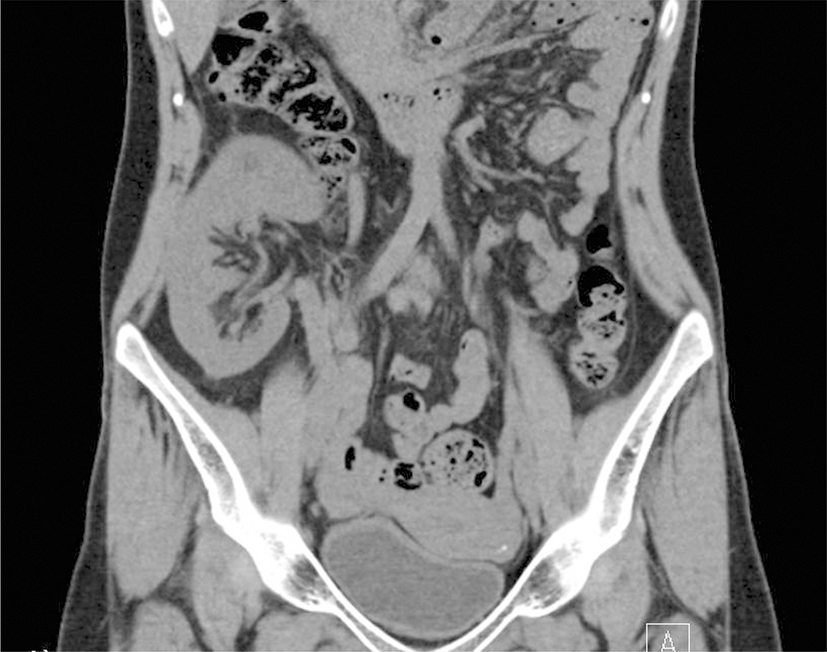
환자는 30세 여자 환자로 만성사구체신염에 의한 말기콩팥병으로 8년간 복막투석을 받아 오다 내원 9개월 전 뇌사자 신이식을 받았다. 수술 후 7일째 시행한 복부초음파에서 저항성지수가 약간 상승된 것 이외 이상 소견 보이지 않았고, 혈중 크레아티닌 0.9 mg/dL로 정상화 되었으며 이후 특별한 증상 없는 상태로 면역억제제 prednisolone, tacrolimus, mycophenolate mofetil을 복용하며 외래에서 관찰 중이었다. 수술 후 6개월 째 복막도관 제거 수술을 시행하였고, 당시 혈중 크레아티닌 0.9 mg/dL였고, 소변검사에서 약간의 농뇨가 관찰되었으나 배뇨증상은 없었으며, 단순복부촬영에서 복막도관 이외 석회화나 비정상적인 음영은 확인되지 않았다. 환자는 내원 4일 전까지 별다른 증상 없이 외래를 방문하였으나 이후 내원 당일 발열, 전신통 및 배뇨통이 발생하여 응급실 통해 입원하였다. 내원 당시 혈압은 120/80 mmHg, 맥박수는 분당 95회, 체온은 38.2°C였으며, 이식신 부위를 촉진하였을 때 압통은 없었고, 오른쪽 늑골척주각 압통이 동반되어 있었다. 일반 혈액검사에서 혈색소 13.2 g/dL, 백혈구 8,000/μL, 혈소판 139,000/μL이었고, 혈중 크레아티닌 1.0 mg/dL, CRP (C-reactive protein)는 10.8 mg/L이었다. 환자의 혈중 칼슘은 8.7 mg/dL, 인산 2.2 mg/dL, 요산 4.7 mg/dL, intact parathyroid hormone은 41.43 pg/mL로 모두 정상이었다. 소변 검사에서는 pH 5.5, 요단백(-), 요잠혈(-), 백혈구(1+), 아질산염 양성 소견을 보였고 고배율 시야에서 30개 이상의 백혈구와 세균이 관찰되었다. 환자는 급성 신우신염으로 진단 받고 ceftriaxone 정주로 치료 받은 후 입원 2일째부터 발열이 호전되었고 옆구리 통증도 지속되긴 하였으나 다소 호전된 상태였으며, 자가 배뇨도 잘 유지되는 상태였다. 입원 3일째 시행한 단순복부촬영에서 방사선 불투과성의 음영이 의심되어(Fig. 1A), 복부초음파를 시행하였고, 이식신의 수신증과 말단 수뇨관을 따라 여러 개의 요관 결석이 발견되었다. 이후 복부 전산화 단층 촬영에서 말단 수뇨관을 따라 쌓여 있는 결석로가 확인되었다(Fig. 1B). 입원 5일째 요관경하 요관결석제거술을 시행하였으나 실패하였고, 당일 오후부터 환자의 소변 양이 감소하여 경피적 신루설치술을 시행하였다. 입원 7일째 개방적 요관결석절제술을 시행하여 결석을 제거하고 이식신의 상단 수뇨관과 본래의 우측 요관 사이 요관요관문합술을 한 후 요관 스텐트를 삽입하였다. 결석의 성분분석 결과 탄산인회석 이었다(Table 1). 24시간 소변검사에서 칼슘과 요산 배설량은 정상이었지만 구연산 수치가 127.6 mg/day (참고범위, 300–900 mg/day)로 감소되어 있어 요중 구연산염 배설을 증가시켜 요로결석 재발을 막기 위해 구연산칼륨을 처방하였다. 2개월 뒤 요관 스텐트와 경피적 신루 카테터를 제거하였고 이후 소변양이 잘 유지되었다. 6개월 뒤 시행한 전산화 단층 촬영에서 요로결석의 재발은 보이지 않았다(Fig. 2).

고 찰

신이식 후 발생하는 요로결석은 흔하지 않은 합병증이다[1-3]. 문헌 고찰에 따르면 신이식 후 요로결석의 발생 빈도는 대개 1–2% 미만으로 보고되고 있다[1,2]. 환자들의 대부분이 이식신의 신경차단으로 통증을 느끼지 못하며, 요로폐쇄에 의한 갑작스런 핍뇨 혹은 무뇨, 혈뇨 등이 종종 통증 없이 발생한다[1,3]. 본 증례에서도 환자가 발열과 배뇨통이 있었지만 이식신 부위 압통을 동반하지는 않았다.

신이식 후 요로결석 형성을 일으키는 소인으로는 고칼슘뇨증, 부갑상선기능항진증, 반복되는 요로감염, 저구연산뇨증, 요관 협착에 의한 요로 폐쇄, 비흡수성 봉합사와 같은 이물질 등이 있다[3,6]. 구연산염은 요로결석 형성을 억제하는 물질로 알려져 있다[7-9]. 따라서 소변에서 구연산염 배설이 감소하면 요로결석 형성을 촉진시킬 수 있다[7,8]. 신이식 환자에서 저구연산뇨증의 빈도가 높다는 보고가 있다[7]. 면역억제제인 cyclosporine A 혹은 tacrolimus와 같은 calcineurin 억제제는 신세뇨관 독성을 일으켜 신세뇨관 산증을 유발할 수 있는데 이 신세뇨관 산증이 소변 구연산염 배설을 감소시킬 수 있는 것으로 알려져 있다[7,9,10]. 본 증례의 환자는 tacrolimus를 지속적으로 복용하던 환자이며, 저구연산뇨증이 동반되어 있었고, 요로 결석 제거 후 구연산칼륨을 지속적으로 투여한 뒤 요로 결석의 재발을 막을 수 있었다. 자발적으로 형성된 요로결석의 경우 결석의 크기가 평균적으로 1.0 cm 정도이고 단일 결석이 대부분이지만[2,3,11] 본 증례의 경우 단일 결석이 아닌 요관 결석로가 발견되었다. 요관 결석로는 전형적으로 체외충격파쇄석술 후 발생하는 합병증이지만[5], 신세뇨관 산증과 연관되어 자발성 결석로가 형성된 사례들이 드물게 보고되어 있다[12,13]. 본 증례는 신이식 후 요관 결석로가 자발적으로 발생한 경우로 저구연산뇨증에 의해 발생하였을 가능성이 높다.

신이식 후 요로결석의 치료는 비신이식 환자에서의 요로결석 치료와 유사하여 결석의 크기 및 위치에 따라 결정된다[2,14]. 크기가 작은 결석을 가진 대부분의 환자들은 보조적 치료를 하면서 경과를 보거나 체외충격파쇄석술로 치료한다[2,3]. 만약 이러한 방법으로 실패할 경우 요관경하배석술 혹은 경피적 신루설치술을 할 수 있다[2,3,14]. 개방적 수술은 거의 필요하지 않고 이식신 주위 섬유화로 시행이 매우 어렵다[14]. 본 증례에선 다수의 요관 결석이 최소침습적 방법으로 제거되지 않았고 대신 개방적 요관결석절제술과 요관요관문합술로 치료에 성공할 수 있었다. 요로결석으로 치료 받은 신이식 환자들 중 추적 관찰 동안 요로결석이 재발하는 사례들이 보고되어 있다[1,2]. 따라서 저구연산뇨증, 고칼슘뇨증, 부갑상선기능항진증과 같은 대사이상이 반드시 함께 교정되어야 한다. 이 외에도 재발을 방지하기 위해서 장기간 추적 관찰이 필요하다.

결론적으로 신이식 후 요로결석은 드문 합병증이지만 요로감염, 요로 폐쇄, 이식신 기능 부전 등을 야기시킬 수 있다. 신이식 환자에서 요로결석은 대부분 전형적인 통증 없이 발생하기 때문에 다양한 상황에서 요로결석의 가능성을 염두에 두는 것이 매우 중요하다. 저구연산뇨증은 신이식 후 흔히 발생하며 요로결석의 위험을 증가시킬 수 있다. 따라서 주의 깊은 감시로 빨리 진단하여 요로결석을 즉시 제거하고 이후에도 재발을 방지하기 위한 예방적 노력이 필요하다.

REFERENCES

1.

Mamarelis G, Vernadakis S, Moris D, Altanis N, Perdikouli M, Stravodimos K, et al. 2014; Lithiasis of the renal allograft, a rare urological complication following renal transplantation: a single-center experience of 2,045 renal transplantations. Transplant Proc. 46:3203-3205

2.

Ferreira Cassini M, Cologna AJ, Ferreira Andrade M, Lima GJ, Medeiros Albuquerque U, Pereira Martins AC, et al. 2012; Lithiasis in 1,313 kidney transplants: incidence, diagnosis, and management. Transplant Proc. 44:2373-2375

3.

Kim H, Cheigh JS, Ham HW. 2001; Urinary stones following renal transplantation. Korean J Intern Med. 16:118-122

4.

Verrier C, Bessede T, Hajj P, Aoubid L, Eschwege P, Benoit G. 2012; Decrease in and management of urolithiasis after kidney transplantation. J Urol. 187:1651-1655

5.

Salem S, Mehrsai A, Zartab H, Shahdadi N, Pourmand G. 2010; Complications and outcomes following extracorporeal shock wave lithotripsy: a prospective study of 3,241 patients. Urol Res. 38:135-142

6.

Harper JM, Samuell CT, Hallson PC, Wood SM, Mansell MA. 1994; Risk factors for calculus formation in patients with renal transplants. Br J Urol. 74:147-150

7.

Stapenhorst L, Sassen R, Beck B, Laube N, Hesse A, Hoppe B. 2005; Hypocitraturia as a risk factor for nephrocalcinosis after kidney transplantation. Pediatr Nephrol. 20:652-656

8.

Hamm LL, Hering-Smith KS. 2002; Pathophysiology of hypocitraturic nephrolithiasis. Endocrinol Metab Clin North Am. 31:885-893

9.

Hamm LL. 1990; Renal handling of citrate. Kidney Int. 38:728-735

10.

Simpson DP. 1983; Citrate excretion: a window on renal metabolism. Am J Physiol. 244:F223-F234

11.

Cicerello E, Merlo F, Mangano M, Cova G, Maccatrozzo L. 2014; Urolithiasis in renal transplantation: diagnosis and management. Arch Ital Urol Androl. 86:257-260

12.

Homayoon K. 1996; Spontaneous steinstrasse due to renal tubular acidosis. Br J Urol. 77:610-611

13.

Van Savage JG, Fried FA. 1993; Bilateral spontaneous steinstrasse and nephrocalcinosi associated with distal renal tubular acidosis. J Urol. 150(2 Pt 1):467-468

14.

Stravodimos KG, Adamis S, Tyritzis S, Georgios Z, Constantinides CA. 2012; Renal transplant lithiasis: analysis of our series and review of the literature. J Endourol. 26:38-44
emj-38-117-f1
Fig. 1. Initial radiologic findings. (A) The plain film of the abdomen reveals radiopaque stones in the pelvic cavity (arrow). (B) Computed tomography scan shows hydroureteronephrosis in transplanted kidney and multiple stones in the distal ureter (steinstrasse).
Download Original Figure
emj-38-117-f2
Fig. 2. Computed tomography after six months. It show no stones in transplanted kidney and ureter.
Download Original Figure
Table 1. Result of Stone analysis
Component %
Calcium oxalate monohydrate Negative
Calcium oxalate dihydrate Negative
Carbonate apatite 100
Uric acid Negative
Brushite Negative
Cysteine Negative
Ammonium urate Negative
Dihydroxyadenine Negative
Protein Negative
Others Negative
Download Excel Table