Introduction
Solitary rectal ulcer syndrome (SRUS) is a rare benign and chronic rectal disease that has a wide spectrum of clinical presentations and variable endoscopic findings [1]. Rectal bleeding and abdominal pain are known as the main symptoms of SRUS. Usually, it is misdiagnosed through colonoscopy [2]; SRUS mimicking rectal cancer according to endoscopic findings, abdominopelvic computed tomography (CT), positron emission tomography (PET)-CT, and magnetic resonance imaging (MRI) has been very rarely reported [3]. We report the case of a 68-year-old man who presented with an ulcerated mass of the rectum representing an SRUS variant.
Case
A 68-year-old man was referred to Jeju National University Hospital with anal pain and difficulty in passing stool. The patient had a history of hypertension and internal hemorrhoids. His vital signs were normal. There was no history of self-digitation, fever, weight loss, or abdominal pain. On initial evaluation, the abdomen was soft, bowel sounds were normal, and there was no tenderness in the abdomen. Rectal examination revealed an irregular broad-based ulcerated mass in the rectum.
Initial laboratory findings were normal (hemoglobin level 16.3 g/dL, white blood cell count 8,900/mm3, and platelet count 182,000/mm3). Additionally, liver function test, erythrocyte sedimentation rate, C-reactive protein, and coagulation profile were normal. Carcinoembryonic antigen (CEA) level was 1.72 ng/mL (normal, 0 to 5.0 ng/mL). Stool occult blood test was negative.
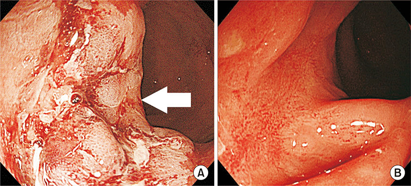
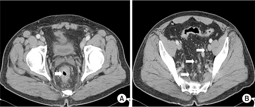
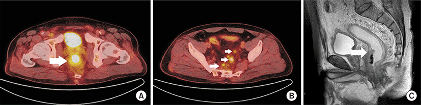
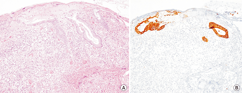
Colonoscopy showed a hemorrhagic and circumferential ulcerated mass with edema in the anterior rectal wall located 5 cm from the anal verge (Fig. 1A). Abdominopelvic CT exhibited an area of rectal wall thickening with perirectal fatty infiltration and enlargement of multiple small mesocolic lymph nodes, consistent with rectal cancer (Fig. 2). However, two repeat biopsies showed ulceration with inflammatory reaction. There was no evidence of malignancy. However, PET-CT was highly suggestive of rectal cancer (maximum standardized uptake value, SUVmax 15.4) with intense fludeoxyglucose uptake in multiple lymph nodes (SUVmax 8.1) (Fig. 3A, B). Furthermore, MRI showed a similar finding, which was irregular wall thickening of mid-rectum with perirectal infiltration (Fig. 3C). Patient underwent repeat biopsy under spinal anesthesia. Incisional biopsy showed a central ulcerated lesion with the surrounding edema invading the muscle layer. A third biopsy also exhibited ulceration with inflammatory reaction (Fig. 4). Two months after conservative management with stool softener and pain control, clinical symptoms of patient improved. Follow-up colonoscopy showed that the lesion markedly improved with remnant ulcerative scarring (Fig. 1B).
Discussion
SRUS is an uncommon disorder of benign and chronic rectal disease with diverse spectrum of clinicopathological abnormalities [4-9]. The mean age at presentation is 30s toith a wide range from 10 to more than 80 years [10]. Although the etiology is presumed to be diverse, the pathogenesis is not entirely understood. First, finger insertion and suppository may cause direct injury [11]. Second, it can be caused by mucosal trauma and ischemia which can be explained by rectal prolapse and paradoxical compression of the pelvic floor [10,12]. There are various symptoms of SRUS [4]. In a single center study, rectal bleeding was present in 82%, abdominal pain in 49%, constipation in 23% and diarrhea in 22%. Histopathological examination is a key to the diagnosis of SRUS. Diagnosis of SRUS is by rule-out of other diseases, ultimately through biopsy. Radiologic examination can be done such as abdominopelvic CT or MRI. However, accurate diagnosis is not always possible, and the treatment is still not established. Since SRUS has a benign disease course, it can be managed with conservative treatment. Patient education and behavioral modification are the first step in the treatment of SRUS, including avoidance of straining and anal digitation, and ingestion of high fiber diet and bulk laxatives. If the symptoms do not improve, mucosal prolapse must be suspected. In the selected patient, biofeedback and surgical treatment may be considered. Using a stool softener can reduce straining during defecation and corticosteroid or mesalazine can also be administered. However, their effectiveness has been not proven. If it cannot be treated medically, surgical treatment may be considered [4].
To date, there have been several reports regarding SRUS [3,9,11,13,14]. Colonoscopic findings with suspected malignancy were variable, including tumor, malignant stricture, and ulcerative mass. MRI was performed frequently as an additional test. In some cases, malignancy and inflammation could not be differentiated. Surgery was performed in several cases. One patient who underwent surgery had intractable symptoms after a short course of ineffective conservative therapy and occult malignancy could not be excluded [15]. In another patient, the ulcer had expanded very quickly to cause a case underwent a second deep biopsy. In most cases, the first biopsy was performed superficially. Some cases underwent a second deep biopsy. Interestingly, they revealed rectal cancer on superficial biopsy initially. However, rectal cancer was ultimately excluded by follow-up circumferential stricture of the rectum [9]. Most cases were treated conservatively, and surgery was avoided [14].
In this case, rectal cancer was strongly suspected in the beginning. On biopsy, only inflammation was revealed. However, confirmation was necessary. The patient did not use finger penetration. Pelvic floor dysfunction and rectal prolapse were not visible. On general diagnostic imaging modalities, neither CT nor MRI had distinguished the inflammation from rectal cancer in a previous report [3]. Even PET-CT was not helpful. Following a number of tests, SRUS was considered, and ultimately diagnosed. In contrast to this case, other cases were initially diagnosed as SRUS and later changed to that of malignancy [14]. Therefore, it is necessary to distinguish carefully for malignancy. Repetitive biopsies are strongly recommended.
In conclusion, SRUS should always be considered in patients with malignant-mimicking rectal cancer. However, we believe it is important not to miss a diagnosis of rectal cancer over the diagnosis of SRUS. We report the case of an SRUS patient who had a central ulcerated mass lesion that mimicked rectal cancer on gross colonoscopic and radiologic findings.