The Ewha Medical Journal
Ewha Womans University School Medicine
Images and Solution

Focal Acral Hyperkeratosis

Ji Yeon Byunhttps://orcid.org/0000-0003-4519-9474, Rosa Kimhttps://orcid.org/0000-0002-0873-7643, Min Young Leehttps://orcid.org/0000-0001-8436-1505, You Won Choihttps://orcid.org/0000-0001-6315-3889, Hae Young Choihttps://orcid.org/0000-0003-3460-2539
Corresponding author Ji Yeon Byun, Department of Dermatology, Ewha Womans University Mokdong Hospital, 1071 Anyangcheon-ro, Yangcheon-gu, Seoul 07985, Korea Tel: 82-2-2650-5159, Fax: 82-2-2652-6925, E-mail: jybyun@ewha.ac.kr

Copyright © 2021 Ewha Womans University School of Medicine. This is an open-access article distributed under the terms of the Creative Commons Attribution Non-Commercial License (http://creativecommons.org/licenses/by-nc/4.0) which permits unrestricted non-commercial use, distribution, and reproduction in any medium, provided the original work is properly cited.

Received: Sep 13, 2021; Revised: Oct 06, 2021; Accepted: Oct 06, 2021

Published Online: Oct 31, 2021


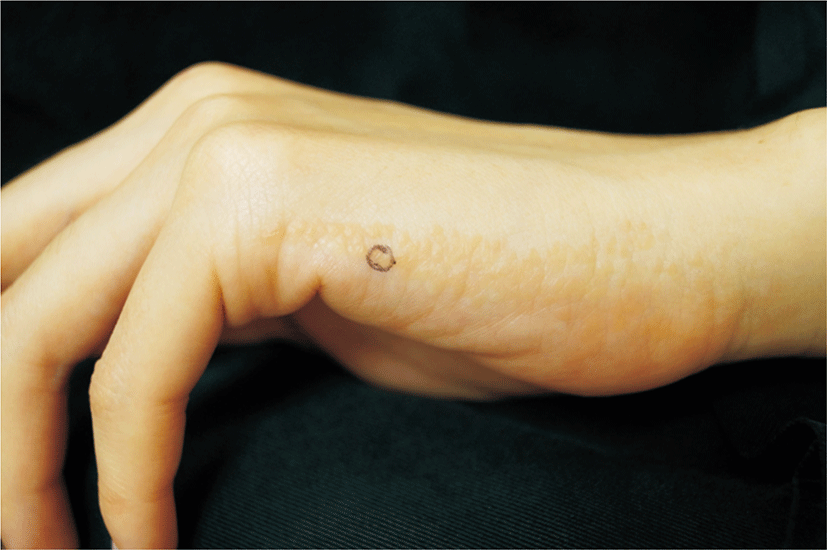
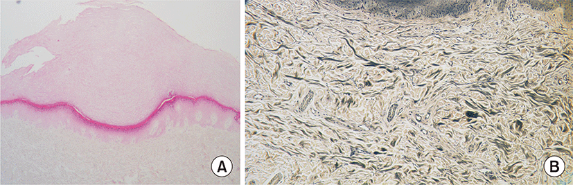
A 29-year-old woman presented with skin lesions on both hands lasted for 1 year. Her past and family history were unremarkable. On skin examination, there were multiple asymptomatic yellow flat discrete papules and plaques along the lateral aspect of both hands (Fig. 1). Skin biopsy revealed focally depressed epidermis with marked hyperkeratosis and hypergranulosis (Fig. 2A). Elastic fiber stain showed normal elastic fibers (Fig. 2B). Based on clinical and histologic findings, the diagnosis of focal acral hyperkeratosis (FAH) was made. The patient was prescribed with topical retinoids and topical keratolytics and showed limited improvement.

FAH is a type of palmoplantar keratoderma, characterized by abnormal thickening of the palms and soles. It occurs both sporadically and as an autosomal dominant genodermatosis. Clinically, multiple skin colored to yellow papules and plaques are localized to the margins of the hands and feet. Histologically, it shows a pronounced hyperkeratosis over a crateriform epidermal depression without dermal change [1,2].

FAH is often considered to be a variant of acrokeratoelastoidosis, although their relationship is controversial. The differential diagnosis with acrokeratoelastoidosis can be made by the absence of elastorrhexis in FAH [1].

Clinically, FAH should be differentiated from verruca vulgaris, but the absence of koilocytes and parakeratosis on histopathologic examination disapproved the possibility of verruca vulgaris in this case.

FAH is a benign condition, yet there is no effective treatment for FAH. Conservative treatment can be tried since this condition is potentially disfiguring. Topical keratolytics, topical retinoids and systemic retinoids are prescribed to decrease hyperkeratosis and relieve symptoms [2,3].

REFERENCES

1.

Samuelov L, Sprecher E. In: Kang S, Amagai M, Bruckner AL, Enk AH, Margolis DJ, McMichael AJ, editors. 2019; Inherited palmoplantar keratodermas. Fitzpatrick's dermatology. 9th edMcGraw-Hill. New York: p. 844-847.

2.

Lee EA, Kim HS, Kim HO, Park YM. 2009; A case of focal acral hyperkeratosis. Ann Dermatol. 21:426-428

3.

Sardana K, Chugh S, Garg VK. 2013; Focal acral hyperkeratosis. Indian Pediatr. 50:256
emj-44-4-144-f1
Fig. 1. Clinical findings. Multiple yellow flat discrete papules and plaques were observed along the lateral aspect of both hands.
Download Original Figure
emj-44-4-144-f2
Fig. 2. Histologic findings. (A) Focal depression of epidermis is observed with marked hyperkeratosis and hypergranulosis (H&E, ×40). (B) There is no alteration of elastic fibers (Verhoeff-van Gieson, ×100).
Download Original Figure