
Introduction
Paget’s disease is a rare intraepithelial neoplasm. It has two sub-types, one is mammary Paget’s disease (MPD) and the other is extramammary Paget’s disease (EMPD) which occurs in other than mammary area. Morphology and histology of these two subtypes are the same. The origin of EMPD is not clear but suggested that they are originated form apocrine bearing glands, such as the perianal region, vulva, scrotum, penis and axilla. The clinical features of EMPD can be non-specific such as chronic eczematous cutaneous disease, therefore misdiagnosis as an inflammatory or infective skin condition is common. Its final diagnosis is often delayed for a long time. The prognosis of this disease entity is variable according to clinical situation and recent analysis of 2,000 EMPD patients showed that EMPD in scrotal lesion, concurrent malignancy, metastatic lesion, old age and male are the risk factors for mortality [1–4]. Chan et al. reported that 8.3% of EMPD patients showed concurrent malignancy in colon and prostate [5]. Sometimes, there are co-existing squamous cell carcinoma in situ in the skin [6].
Surgical resection with negative margin is the considered to be the standard of care in EMPD, other options include imiquimod 5% topical cream, modified peripheral Mohs surgery and radiation therapy [7,8]. Although recurrence is common, the disease has a slow progression and paucity metastasis, so the prognosis is generally good.
Here, we describe a case of rapid progressed EMPD patient, who showed fulminant disease course in two months.
Case Report
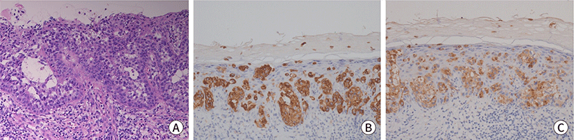
A 70-year-old man presented with discomfort in the right inguinal area. He had a history of skin malignancy in the right inguinal area three years ago and treated with surgical resection (wide excision and split thickness skin graft from thigh). The histopathological study at that time showed mostly squamous cell carcinoma (3×2.5 cm sized with depth of 4 mm invasion) and focal EMPD component (Fig. 1). During the last three years, he did not visit the clinic. This time he visited the hospital due to right inguinal discomfort. In physical examination, he showed palpable right inguinal lumps without obvious skin change. CT scan showed difuse skin thickening and subcutaneous infiltration at right inguinal and pubic area with mild lymph node enlargements. There was no other visceral lesion in the CT scan (Figs. 2A, 2B). Blood test showed no specific finding except for a mild CEA elevation (10.4 ng/mL, reference range 0–5.0 ng/mL). He underwent a punch biopsy in right inguinal lymph nodes area. The pathology showed the consistent findings of EMPD and immunohistochemical stains showed CK7 (+), CEA (+) and HER2 SISH (–). Further groin dissection was planned. However, he wanted to postpone surgery due to private reasons.
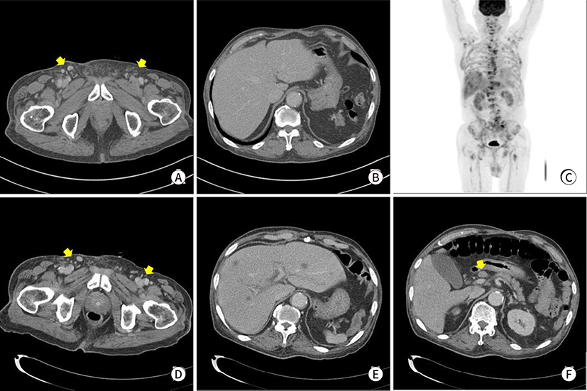
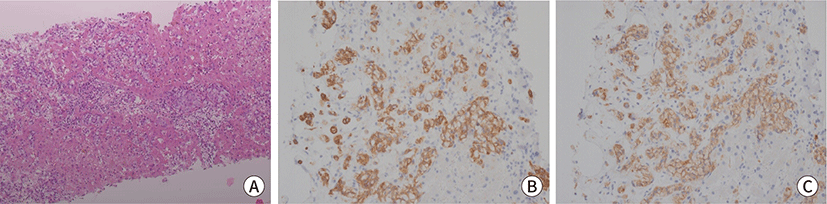
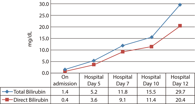
However, two months later, he visited the clinic again due to severe back pain. Spine MRI showed several metastatic lesions in the lumbar spine, sacrum and pelvic bones. The blood test showed anemia with hemoglobin level of 6.1 g/dL and elevated CEA level (21.2 ng/mL) compared with results obtained two months ago. Endoscopic findings including colonoscopy and duodenofiberscopy showed no bleeding tendency or no specific lesion. Subsequent CT scan showed multiple hepatic metastasis, various lymph nodes enlargements, and bony metastasis in abdomen. However no significant change of inguinal lymphadenopathies was documented as EMPD two months ago. FDG PET scan showed similar findings (Figs. 2C–2F). At the time of admission, the level of bilirubin was in normal range, but suddenly he showed jaundice with increasing serum bilirubin level. He underwent liver biopsy and the pathologic results showed mmetastatic carcinoma from the previous EMPD with the same immunohistochemical results of CK7 (+), CEA (+), GCDFP-15(+), HER2 (–) (Fig. 3). We planned chemotherapy for the metastatic EMPD. However just a few days, he showed rapid clinical deterioration with hepatic failure (Fig. 4). Eventually, on the 12nd admission day, he died of the disease.
This is the first report about a fulminant disease course of a metastatic, recurrent EMPD to this time in our country.
Discussion
EMPD is a rare slow-growing cutaneous adenocarcinoma that presents as an erythematous, eczematous plaque outside the mammary gland. This neoplastic lesion is often delayed before diagnosis, but usually resulted good prognosis. The range of EMPD recurrence is very wide, for the example, the recurrence of EMPD in vulva varies from 12% to 61% [5,9]. On multiple variate analysis, dermal invasion, lymph node metastasis and elevated serum CEA associated with poor prognosis have been reported [10]. The analysis from large scale patients showed that surgery is a protective factor and radiation is a risk factor for EMPD survival [4]. Although the invasion of dermis is uncommon, if once the EMPD invades into the dermis, that could get metastatic characteristics [11]. Interestingly, Chanda et al. reported that 46% of patients with EMPD associated with an underlying carcinoma developed metastatic disease, compared to 18% of patients with a non-invasive disease [12]. In a large study of 1,439 patients with invasive EMPD, disease specific five year survival was 94.9% (95% CI 92.7%–96.5%) for localized disease, 84.9% (95% CI 77.4%–90.0%) for regional disease and 52.5% (95% CI 29.3%–71.3%) for distant disease. For patients diagnosed with distant spread the outlook was still favorable since more than half of all patients were alive at 5 years of observation [13].
In the case of our patient, he had the risk factor of concurrent skin squamous cell carcinoma, his EMPD presented just as a minor component of that skin squamous cell carcinoma. However, after relapse, EMPD was the only lesion except squamous cell carcinoma. Meanwhile, the most interesting things in this case is that he rapidly progressed to death within three months after diagnosis of relapse. Actually this patient visited EMPD recurrence after 3 years of primary treatment, and there was an evidence of dermal invasion such as subcutaneous infiltrative lesions in the CT scan without visceral or distant metastasis. However, two month’s break resulted in distant failure and finally death. Thus, the findings of regional node involvement mimicking lymphadenitis would be a grave sign for rapid distant progression. Recently one study has shown that systemic treatment of EMPD in the disease is metastatic, especially some EMPD showed HER2 positivity and anti-HER2 therapy could result in a durable response and durable survival [14].
It is still uncertain whether long standing local recurrent disease will transform to invasive form. Chang et al. reported that chemokine receptors, CXCR4 and CXCR7 are associated with poor outcome and that they can be used as prognostic biomarkers [15]. In the recent basic and translational research, many other disease mechanisms are under elucidation including RAS-RAF-MEK-ERK signaling, PI3K-AKT-mTOR signaling and androgen-AR signaling. In the future, diagnostic and therapeutic application of this research outcome would make the management of EMPD much better.
Conclusion
EMPD is an intraepithelial malignancy, which shows recurrence and occasional metastasis, with usually good prognosis. Although several studies reported this disease entity, the focus was on the initial skin lesions with limited experience of disease locations and surgical outcomes. This study is the first report of a poor clinical outcome in three months of presentation involving aggressive deterioration of metastatic EMPD. The findings aid physicians in selectively managing EMPD patients, especially those diagnosed with the disease concomitant with squamous cell carcinoma of skin, for enhanced clinical out-comes.