The Ewha Medical Journal
Ewha Womans University School Medicine
Case Report

죽상판 없는 우관상동맥의 심한 음성 혈관 재형성 1예

장경윤, 박경선, 최유아, 김지희, 전부석, 허성호
Kyung Yoon Chang, Kyung Seon Park, Yoo A Choi, Ji Hee Kim, Bu Seok Jeon, Sung Ho Her
Corresponding author: Sung Ho Her, Division of Cardiology, Department of Internal Medicine, Daejeon St. Mary’s Hospital, The Catholic University of Korea School of Medicine, 64 Daeheung-ro, Jung-gu, Daejeon 301-723, Korea Tel: 82-42-220-9504, Fax: 82-42-253-9505E-mail: hhhsungho@catholic.ac.kr

Copyright © 2012 Ewha Womans University School of Medicine. This is an open-access article distributed under the terms of the Creative Commons Attribution Non-Commercial License (http://creativecommons.org/licenses/by-nc/4.0) which permits unrestricted non-commercial use, distribution, and reproduction in any medium, provided the original work is properly cited.

Received: Jan 20, 2012; Accepted: Feb 28, 2012

Published Online: Mar 31, 2012

Abstract

Arterial remodeling is commonly observed in human atherosclerosis. It is a heterogeneous response ranging from positive remodeling to negative remodeling. Negative remodeling is a condition in which the vessel area decreases in size, often as a result of a structural change in the coronary vessel wall. But its contribution to myocardial ischemia in a de novo lesion has not been clearly shown. A 51-year-old female with exertional angina was admitted to our hospital. Coronary angiography was performed, revealing a severe stenosis at the middle part of the right coronary artery (RCA). Although we predilated ballooning at the middle RCA, the degree of stenosis did not improve. Thus intravascular ultrasound (IVUS) was performed. The lesion was not nearly showed plaque burden and severe negative remodeling. Though the cross-sectional narrowing percentage was significant, we decided to medical treatment for fearing coronary perforation by stenting. This case report intends to emphasize that severe coronary stenosis should be performed IVUS before the stenting. We describe a rare case with severe negative remodeling at the middle part of the RCA without atheroma plaque.

Keywords: Angina; Atherosclerosis; Ultrasound, intravascular; Negative remodeling

서 론

혈관 재형성은 동맥 경화증 환자에서 흔히 관찰된다. 급성 관상동맥 증후군 환자에서 병변 부위와 관련되어 혈관 직경이 증가하는 양성 재형성부터 만성 안정형 협심증 환자에서 혈역학적으로 의미 있는 협착을 동반하여 혈관 직경이 감소하는 음성 재형성까지 다양하게 나타난다[1-5].

특히, 음성 재형성은 관상동맥혈관 벽의 구조적 변화의 결과로 혈관의 내경이 좁아지는 것을 말한다. 이는 풍선 혈관 확장술 시행 후 재 협착의 중요한 인자가 되나 심근경색 환자에서 새롭게 생기는 경우는 흔하지 않다[6-8]. 저자들은 운동성 흉통을 호소하는 환자에서 시행한 관상동맥 혈관 조영술에서 우관상동맥 중간부위의 죽상판 없는 심한 음성 혈관 재형성이 관찰되어 약물로 성공적으로 치료하였기에 문헌고찰과 함께 보고하는 바이다.

증 례

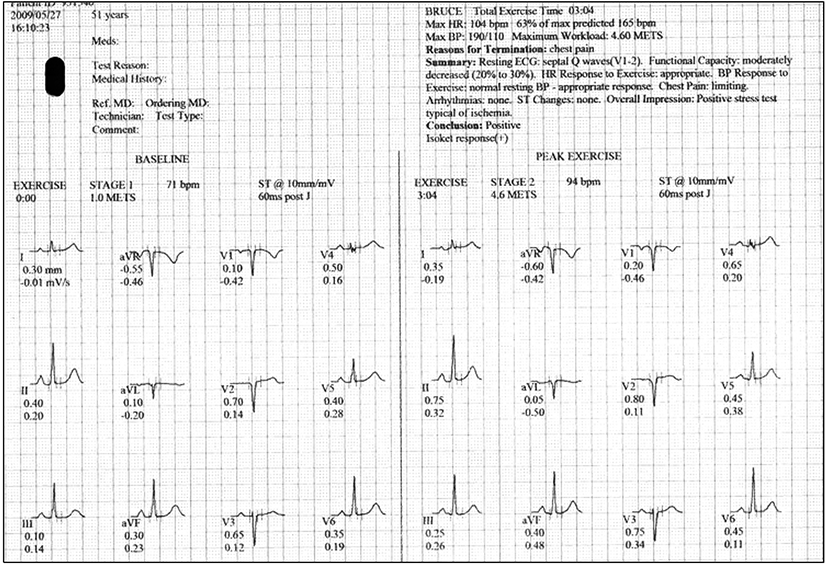
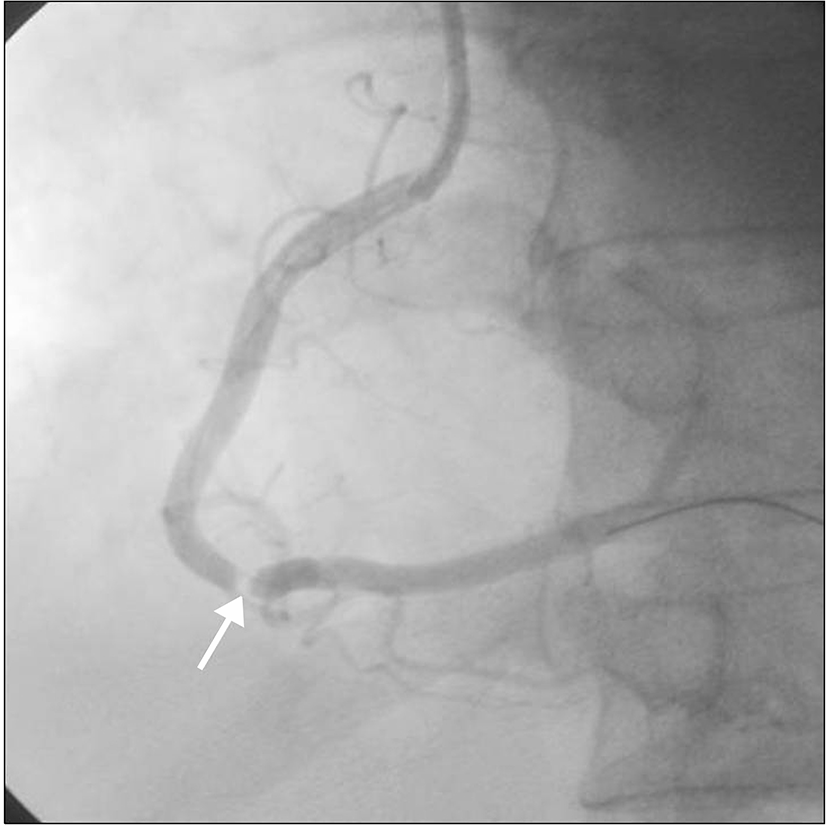
51세 여자가 내원 전일부터 발생한 운동성 흉통을 주소로 내원하였다. 비흡연자로 과거력 및 가족력상 특이소견은 없었다. 내원 당시 활력 징후는 혈압 110/ 70 mmHg, 맥박 72회/분, 호흡수 16회/분, 체온 36.5도였다. 흉부진찰에서 호흡음 및 심음은 이상이 없었다. 심전도는 정상 동율동, ST분절 이상은 없었다. 운동부하 심전도 검사에서 4.6 METs에서 흉통이 발생하였고 양성 소견을 보였다(Fig. 1). 심장 관류 단일광자 단층 촬영(Adenosine Technetium-99m [Tc-99m] sestamibi myocardial perfusion single photon emission computed tomography, SPECT)에서 스트레스시 우측 관상동맥 영역에서 심근 경색 소견이 관찰되었다. 심혈관 관상동맥 혈관 조영술을 시행하였고 우관상동맥의 중간부위에 심한 협착이 관찰되었다(Fig. 2).

혈관 연축을 감별 하기 위해 우관상동맥으로 nitroglycerin 200 mg를 주입하였고, nitroglycerin 정주하였으며, 우관상동맥에 2.75×20 mm 풍선 확장술을 시행하였다. 하지만 협착

정도는 호전되지 않았다. 확장되지 않는 관상동맥의 석회화 여부 및 동맥경화증 진행 정도를 확인하기 위해 혈관 내 초음파(iLab, Boston Scientific, Natick, MA, USA)를 시행하였다. 혈관 내 초음파 도자를 생리식염수로 세척한 후 유도선을 따라 병변 원위부까지 통과시킨 후 천천히 다시 반대방향으로 병변을 지나 병변 근위부 표준혈관까지 혈관 내 초음파 도자를 이동시키면서 영상을 얻었다. 이를 통해 외측 탄력막(external elastic membrane, EEM) 면적을 측정하였다. 흥미롭게도 죽상판은 관찰되지 않았으며, 심한 음성 혈관 재형성 소견이 관찰되었다. 협착부위 외측 탄력막 면적은 2.27 mm2였으며 병변 말단부위의 외측 탄력막은 13.2 mm2로 그 비율은 0.17이었다(Fig. 3). 우관상동맥의

음성 혈관 재형성에 대해 단면 협착비율은 의미 있는 수치였음에도 불구하고, 스텐트 삽입술시 관상동맥 파열 등의 합병증 발생 가능성을 고려하여 약물치료를 하기로 하였다. Bisoprolol (Concor 2.5 mg; Merck, Darmstadt, Germany)과 nicorandil (Sigmart 5 mg bid; JW Pharmaceutical, Seoul, Korea)을 추가하였고 흉통은 호전되었다. 현재 약물 유지하면서 1년째 외래에서 추적관찰 중이다.

고 찰

관상동맥 혈관 재형성은 Glagov 등[3]에 의해 처음 기술되었다. 혈관 직경은 실질적으로 단면적이 40%이상 감소시 내경이 좁아지는데, 이는 내측 탄력 섬유막(internal elastic lamina)의 보상적 확장 때문이다. Clarkson 등은 부검을 통해 동맥 경화성 좌전하행동맥을 관찰하였을 때 내경의 크기가 협착부위의 단면적에 독립적임을 밝혔다[9-11].

이전 여러 연구에서 혈관 내 초음파를 통해 세 관상동맥의 새로운 병변의 혈관 재형성 평가를 보고한 바가 있다[12]. 외측 탄력막 면적의 감소가 우관상동맥보다 좌전하행 동맥이나 좌선회동맥에서 많은데, 좌전하행동맥이나 좌선회동맥이 주분지 혈관 근처에 위치하기 때문에 이런 외측 탄력막 감소 및 주분지 혈관이 혈관 재형성을 평가하는데 영향을 미치게 된다[13]. 여러 연구에서 혈관재형성에 영향을 미치는 전신적 인자에 대한 보고들이 있었다. 주로 양성 재형성은 고콜레스테롤혈증[14], 급성 관상동맥 증후군에서 흔하며[4], 음성 혈관재형성은 흡연자와 인슐린을 사용하는 당뇨환자에서 더 많았다[5,15].

양성 혈관재형성을 연구한 체내 혈관 내 초음파 연구는 증가했지만, 음성 혈관재형성에 대한 연구는 제한적이었고 뿐만 아니라 음성 재형성의 증례보고 역시 기시부에 국한되어 있었다(Table 1) [1,16]. Kobayashi 등[16]은 좌선회동맥 근위부에서 60% 협착 있는 경우에서 혈관 내 초음파를 시행하여 관찰된 경한 죽상판과 음성 재형성에 대해 스텐트 삽입시 발생할 수 있는 관상동맥 파열에 대해 우려하여 약물치료를 하였다. 반면 Sadamatsu 등[6]은 음성 재형성이 있는 환자에서 의미 있는 죽상판이 없는 근위부 병변에 스텐트 삽입술을 시행하였는데, 차후 심근경색의 주원인이 되었다.

최근 한 보고에 의하면 혈관 내 초음파를 연속적으로 사용하여 정상 부분과 병변 부위 모두 외측 탄력막 면적의 증가여부를 관찰하였는데, 18개월 추적관찰 하였을 때 병변 부위 보다 정상부분에서 증가 폭이 더욱 컸다[3]. 이런 증가는 관상동맥 병변 부위의 광범위한 재형성을 나타낸다. 주로 관상동맥의 기시부에서 음성 재형성이 관찰되는데, Shimada 등[5]은 좌전하행동맥 주분지의 직하방 분지점에서 음성 재형성 증례를 보고하였다. 본 증례는 혈관 내 초음파를 통해 죽상판 없이 우관상동맥의 중간부분에서 심한 음성 혈관 재형성이 관찰된 드문 예로 약물 치료로 성공적으로 치료하였기에 문헌고찰과 함께 보고하는 바이다.
REFERENCES

1.

Nishioka T, Luo H, Eigler NL, Berglund H, Kim CJ, Siegel RJ. 1996; Contribution of inadequate compensatory enlargement to development of human coronary artery stenosis: an in vivo intravascular ultrasound study. J Am Coll Cardiol. 27:1571-1576

2.

Mintz GS, Kent KM, Pichard AD, Satler LF, Popma JJ, Leon MB. 1997; Contribution of inadequate arterial remodeling to the development of focal coronary artery stenoses: an intravascular ultrasound study. Circulation. 95:1791-1798

3.

Glagov S, Weisenberg E, Zarins CK, Stankunavicius R, Kolettis GJ. 1987; Compensatory enlargement of human atherosclerotic coronary arteries. N Engl J Med. 316:1371-1375

4.

Schoenhagen P, Ziada KM, Kapadia SR, Crowe TD, Nissen SE, Tuzcu EM. 2000; Extent and direction of arterial remodeling in stable versus unstable coronary syndromes : an intravascular ultrasound study. Circulation. 101:598-603

5.

Shimada Y, Courtney BK, Nakamura M, Hongo Y, Sonoda S, Hassan AH, et al. 2006; Intravascular ultrasonic analysis of atherosclerotic vessel remodeling and plaque distribution of stenotic left anterior descending coronary arterial bifurcation lesions upstream and downstream of the side branch. Am J Cardiol. 98:193-196

6.

Sadamatsu K, Maehira N, Tashiro H. 2007; Negative remodeling at the ostium of the left anterior descending artery induced myocardial ischemia. J Invasive Cardiol. 19:E328-E330.

7.

Wexberg P, Gyongyosi M, Sperker W, Kiss K, Yang P, Hassan A, et al. 2000; Pre-existing arterial remodeling is associated with in-hospital and late adverse cardiac events after coronary interventions in patients with stable angina pectoris. J Am Coll Cardiol. 36:1860-1869

8.

Okura H, Hayase M, Shimodozono S, Bonneau HN, Yock PG, Fitzgerald PJ. 2001; Impact of pre-interventional arterial remodeling on subsequent vessel behavior after balloon angioplasty: a serial intravascular ultrasound study. J Am Coll Cardiol. 38:2001-2005

9.

Clarkson TB, Prichard RW, Morgan TM, Petrick GS, Klein KP. 1994; Remodeling of coronary arteries in human and nonhuman primates. JAMA. 271:289-294

10.

Sipahi I, Tuzcu EM, Schoenhagen P, Nicholls SJ, Crowe T, Kapadia S, et al. 2006; Static and serial assessments of coronary arterial remodeling are discordant: an intravascular ultrasound analysis from the Reversal of Atherosclerosis with Aggressive Lipid Lowering (REVERSAL) trial. Am Heart J. 152:544-550

11.

Burke AP, Kolodgie FD, Farb A, Weber D, Virmani R. 2002; Morphological predictors of arterial remodeling in coronary atherosclerosis. Circulation. 105:297-303

12.

Hong MK, Mintz GS, Lee CW, Kim YH, Lee JW, Song JM, et al. 2003; Intravascular ultrasound assessment of patterns of arterial remodeling in the absence of significant reference segment plaque burden in patients with coronary artery disease. J Am Coll Cardiol. 42:806-810

13.

Javier SP, Mintz GS, Popma JJ, Pichard AD, Kent KM, Satler LF, et al. 1995; Intravascular ultrasound assessment of the magnitude and mechanism of coronary artery and lumen tapering. Am J Cardiol. 75:177-180

14.

Tauth J, Pinnow E, Sullebarger JT, Basta L, Gursoy S, Lindsay J, et al. 1997; Predictors of coronary arterial remodeling patterns in patients with myocardial ischemia. Am J Cardiol. 80:1352-1355

15.

Kornowski R, Mintz GS, Lansky AJ, Hong MK, Kent KM, Pichard AD, et al. 1998; Paradoxic decreases in atherosclerotic plaque mass in insulin-treated diabetic patients. Am J Cardiol. 81:1298-1304

16.

Kobayashi Y, Mehran R, Moussa I, Reyes A, Moses JW. 2001; Negative remodeling at the ostium of the left circumflex artery. J Invasive Cardiol. 13:805-807.
emj-35-065-f1
Fig. 1. Exercise treadmill test was positive by typical angina at 4.6 METs.
Download Original Figure
emj-35-065-f2
Fig. 2. Right coronary angiogram revealed a severe stenosis at the middle part of the right coronary artery (arrow).
Download Original Figure
emj-35-065-f3
Fig. 3. Intravascular ultrasound was showed severe negative remodeling (B) without atheroma plaque compared to the proximal and distal site (A, C).
Download Original Figure
Table 1. Cases of negative remodeling
Disease Method Lesion Treatment Result References
Stable angina IVUS LCx ostium Medical Improved Kobayashi et al. [16]
Stable angina IVUS LAD ostium Intervention MI Sadamatsu et al. [6]
Stable angina IVUS Mid RCA Medical Improved This study

IVUS, intravascular ultrasound; LCx, left circumflex artery; LAD, left anterior descending artery; RCA, right coronary artery; MI, myocardial infarction.

Download Excel Table