Introduction
Pancreatic panniculitis is a rare skin complication of subcutaneous fat necrosis that develops in 0.3% to 3% of patients with pancreatic disease [1]. Most patients present with tender red-brown nodules on the lower extremities. The lesions may spontaneously exude an oily material due to the liquefactive necrosis of adipocytes.
The diagnosis is based on the presence of a pancreatic disorder as determined by laboratory tests, imaging, and skin biopsy. The main histologic features are lobular fat necrosis, anuclear adipocytes with a thick, shadowy wall (ghost cells), and focal calcification with a mixed inflammatory infiltrate [2].
Here we report a rare case of pancreatic panniculitis in a patient with acute pancreatitis and a review of the related literature.
Case
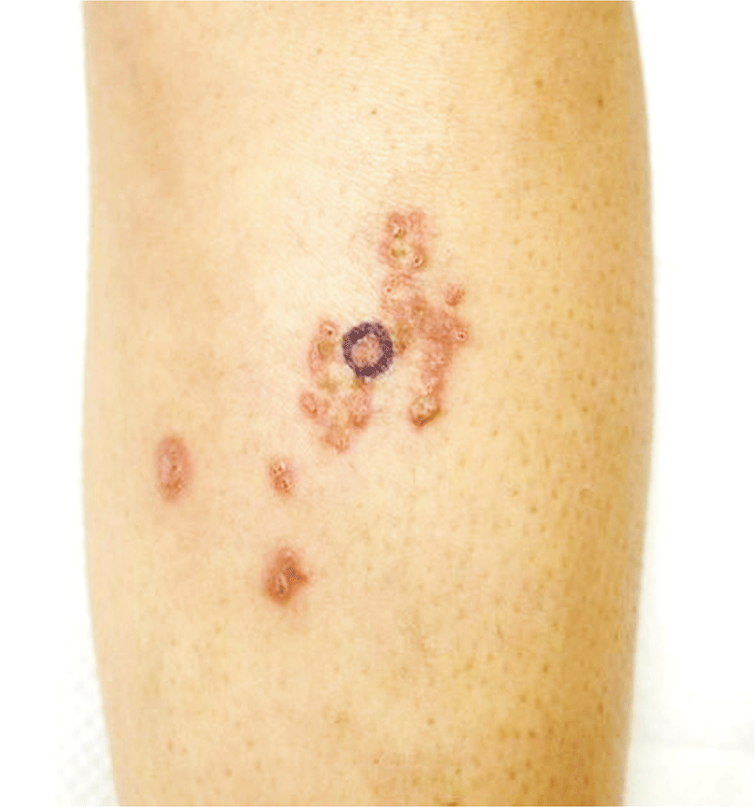
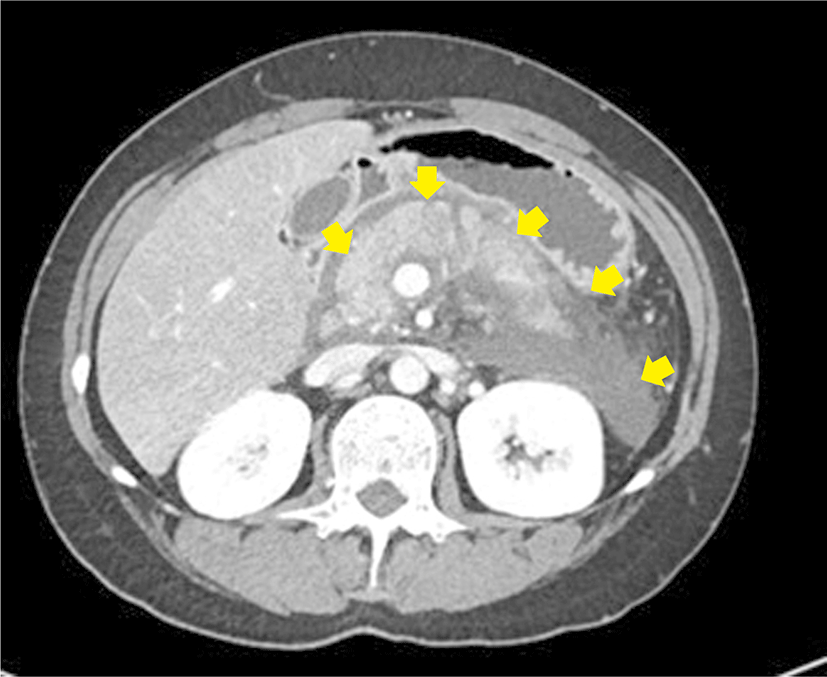
A 32-year-old female was referred to our clinic for multiple tender nodules with erythematous crusts on her left shin (Fig. 1). She had a history of alcoholic liver cirrhosis and had been hospitalized multiple times for acute pancreatitis secondary to alcohol abuse. On admission to our clinic, she had acute abdominal pain, nausea, and vomiting. The blood examination revealed an increased leukocyte count (16,540/mm3; normal, 4,000–10,000/mm3), an elevated C-reactive protein level (7.45 mg/dL; normal, 0–0.5 mg/dL), and significantly elevated serum amylase (434 IU/L; normal, 28–100 IU/L) and serum lipase (2,055 U/L; normal, 13–60 U/L) levels. Computed tomography of the abdomen revealed an increased pancreatic volume and peripancreatic infiltration extending from the head to the tail of the pancreas, consistent with a diagnosis of acute pancreatitis (Fig. 2). She was started on intravenous fluids, antibiotics, and analgesics.
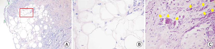
A physical examination performed at that time revealed multiple crusted red-brown subcutaneous nodules on her left lower extremity. The histopathological findings of a skin biopsy taken from a nodule included lobular panniculitis, without any signs of vasculitis, as well as diffuse fat necrosis. The latter consisted of anucleated adipocytes with shadowy cell membranes. Fine basophilic calcium deposits were present in the dermis and subcutaneous fat (Fig. 3). These findings were consistent with a diagnosis of pancreatic panniculitis.
The patient was discharged after receiving conservative treatment for acute pancreatitis. However, she continued to consume alcohol, resulting in repeated hospitalizations due to recurrent pancreatitis. She received an intralesional injection of triamcinolone acetonide for the management of the panniculitis, but this resulted in only minimal improvement of the skin lesions, which recurred with repeated exacerbations of her pancreatic disease.
Discussion
Pancreatic panniculitis was first described by Chiari in 1883 and has since been reported in conjunction with other pancreatic pathologies, such as acute and chronic pancreatitis, pancreatic cancer, and pancreatic pseudocysts [3]. The exact etiology of pancreatic panniculitis is unknown, but the features of the disease include the release of trypsin and other pancreatic enzymes as well as increased microcirculatory permeability, with subsequent hydrolysis of neutral fat by lipase and amylase [4]. The resultant release of glycerol and free fatty acids leads to necrosis of the adipocytes and accompanying inflammatory responses. The released free fatty acids combine with calcium to form basophilic deposits. A pathogenic role of the pancreatic enzymes is supported by elevated levels of lipase and anti-lipase monoclonal antibodies within the necrotic subcutaneous tissue [5]. However, some patients with pancreatic panniculitis have normal pancreatic enzyme levels, suggesting a role for unrecognized factors in the development of the disease [4].
The histopathological hallmark of pancreatic panniculitis is the presence of “ghost cells,” i.e., anucleated adipocytes with shadowy thick cell membranes. The main histologic pattern is mostly lobular panniculitis without vasculitis, but a septal pattern of panniculitis may be seen in the very early stage of the disease [5]. Fat hydrolysis followed by a reaction with calcium results in the formation of granular basophilic deposits within the cells, such that they stain positively with von Kossa stain.
The differential diagnosis of subcutaneous nodules in a patient with pancreatic panniculitis include other forms of panniculitis, such as alpha-1-antitrypsin deficiency panniculitis, erythema nodosum, erythema induratum, and polyarteritis nodosa. Skin biopsy is often needed to distinguish these conditions. Erythema nodosum can be differentiated based on its septal panniculitis pattern, seen on histopathologic examination. Patients with erythema induratum and polyarteritis nodosa typically present with vasculitis in addition to panniculitis. The histologic features of alpha-1-antitrypsin deficiency panniculitis resemble those of lobular panniculitis with neutrophil infiltration, but extensive enzymatic fat necrosis with saponification and calcium deposits is lacking [6].
The treatment of pancreatic panniculitis is mainly supportive and depends primarily on managing the underlying pancreatic disease. In acute pancreatitis, the skin lesions usually resolve when the inflammation subsides. In patients with pancreatic cancer, the skin lesions tend to be more persistent, requiring surgical excision of the tumor followed by resolution of the skin lesions in some patients [7]. In our patient, the underlying necrotizing pancreatitis recurred, further aggravating the chronic disease course, such that the skin lesions did not show remarkable improvement.
In summary, pancreatic panniculitis is a rare skin manifestation of subcutaneous fat necrosis. To the best of our knowledge, only 10 cases have been reported in the Korean literature [8].