

| Won-joong Kim | 3 Articles |
|
[English]
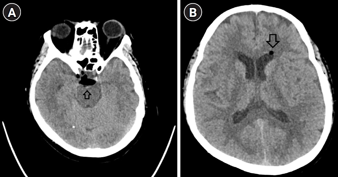
Pneumocephalus is a rare complication of neuraxial procedures and is usually associated with inadvertent dural puncture or the use of air during epidural space identification. Epiduroscopic epidural neuroplasty (EEN) is performed without air injection and permits direct visualization of the epidural space; therefore, pneumocephalus after this procedure is extremely uncommon. A 71-year-old woman with a history of lumbar spine surgery underwent EEN via a caudal approach without sedation. No dural puncture was identified during the procedure. Approximately 12 hours later, she developed a non-orthostatic headache. Brain computed tomography revealed pneumocephalus in the basal cistern and left lateral ventricle. Her symptoms improved with oxygen therapy but worsened 5 days later despite radiologic resolution of the pneumocephalus. Suspected cerebrospinal fluid (CSF) leakage was treated with an epidural blood patch, which resolved her symptoms. Pneumocephalus may occur after EEN without clinically or endoscopically recognized dural injury and may coexist with CSF leakage; an occult microdural defect cannot be excluded as the underlying mechanism. Prompt imaging should be considered in patients who develop early or atypical postprocedural headache.
[English]
Although sciatica is commonly associated with lumbar spinal issues, it is important to acknowledge that non-spinal factors can also play a significant role in this condition. This is particularly relevant for female patients, in whom gynecologic conditions can lead to secondary sciatic neuropathy. Herein, we report the case of a 66-year-old woman who experienced posterolateral right lower extremity radiating pain. We initially performed a lumbar transforaminal epidural steroid injection, but the pain persisted. Subsequently, hip MRI revealed sciatic neuropathy adjacent to the pedunculated portions of a uterine myoma. We then performed a sub-gluteal sciatic nerve block under ultrasound guidance, resulting in significant relief of her pain. In conclusion, hip MRI can be helpful for the differential diagnosis of sciatica, and ultrasound-guided sciatic nerve block can be considered an appropriate and effective treatment option. Citations Citations to this article as recorded by

[English]
An 84-year-old woman visited our pain clinic with complaints of low back pain and severe radiating pain in the right lower extremity during walking. The patient demonstrated subacute compression fracture of L3 with vacuum change in lumbar spine plain radiographs and MRI which suggest Kummell’s disease. Despite our conservative treatments, she had little back pain relief. Therefore, we planned a percutaneous vertebroplasty. Manual compression could help perform percutaneous vertebroplasty more effectively by expanding the vertebral body. In addition, the spontaneous recovery of vacuum cleft width using negative pressure could help perform the technique more effectively. We successfully performed percutaneous vertebroplasty using these combination therapies for our patient.
|
|
 , Bo Kyung Kang