• Contact us
  • E-Submission
ABOUT
BROWSE ARTICLES
JOURNAL POLICIES
FOR CONTRIBUTORS

Page Path

3
results for

"Myocarditis"

Filter

Article category

Keywords

Publication year

Authors

"Myocarditis"

Original article

[English]
Purpose
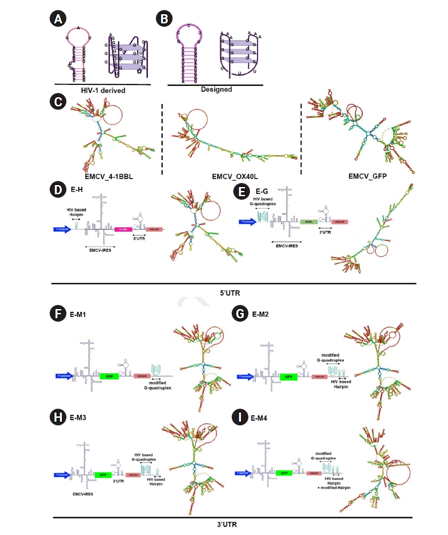
Internal ribosome entry site (IRES) elements, present in both viral and cellular messenger RNAs (mRNAs), facilitate cap-independent translation by recruiting ribosomes to internal regions of mRNA. This study aimed to investigate the impact of inserting G-quadruplex and hairpin structures into the 5' untranslated region (UTR) and poly(A) sequences on the translation efficiency of the encephalomyocarditis virus (EMCV) IRES, using an IRES-based RNA platform encoding OX40L, 4-1BBL, and GFP.
Methods
G-quadruplex and hairpin structures, derived from HIV-1 (human immunodeficiency virus type 1) or custom-designed, were synthesized and inserted into the 5' UTR and poly(A) tail regions of EMCV IRES vectors. These constructs were amplified by polymerase chain reaction, ligated into plasmids, and transcribed in vitro. B16 melanoma, TC-1 tumor, and HEK293 cells were transfected with these RNA constructs. Protein expression levels were assessed at 6, 12, and 24 hours post-transfection by flow cytometry and fluorescence microscopy. Statistical analyses employed one-way analysis of variance with the Dunnett test.
Results
The insertion of G-quadruplex and hairpin structures altered RNA secondary structure, significantly reducing protein expression. In the 5' UTR, the G-quadruplex nearly abolished OX40L expression (1.18%±0.41% at 6 hours vs. 18.23%±0.16% for control), while the hairpin structure reduced it (16.29%±1.46% vs. 22.84%±1.17%). In the poly(A) tail region, both structures decreased GFP expression across all cell lines (4.86%±1.35% to 7.27%±0.32% vs. 39.56%±2.07% in B16 cells).
Conclusion
Inserting G-quadruplex and hairpin structures into EMCV IRES UTRs inhibits translation efficiency, suggesting the need for precise RNA structure modeling to enhance IRES-mediated translation.
  • 209 View
  • 5 Download
Case Reports
[English]
Acute Fulminant Myocarditis Recovered from Electro-Mechanical Dissociation in Scrub Typhus
Young Hak Jung, Minjoo Lee, Kyoung Hwa Lee, Ji Hoon Lee, Seok-hyung Kim, Byoung Kwon Lee
Ewha Med J 2016;39(1):1-5.   Published online January 29, 2016
DOI: https://doi.org/10.12771/emj.2016.39.1.1

Scrub typhus, caused by Orientia tsutsugamushi, is an acute febrile illness. Characteristics of tsutsugamushi disease are fever, rash and eschar. However, severe complications might rarely occur, such as acute fulminant myocarditis caused by scrub typhus. Thus, there are few reports of recovery from seriously complicated cases. We report on an adult male with scrub typhus complicated with acute fulminant myocarditis with no previous comorbid illness who recovered successfully with proper treatment including antibiotics, ventilator support, percutaneous cardiopulmonary support, and continuous renal replacement therapy.

Citations

Citations to this article as recorded by  
  • Comprehensive review on cardiac manifestation of scrub typhus
    Barath Prashanth Sivasubramanian, Abul Hasan Shadali Abdul Khader, Diviya Bharathi Ravikumar, Francis Vino Dominic Savio, Umabalan Thirupathy, Varshini Thiruvadi, Rhea Prasad, Hema Thokala, Husna Qadeer, Dhiraj Poragal Venkataperumal, Ashima Gupta, Nagara
    Frontiers in Tropical Diseases.2024;[Epub]     CrossRef
  • Case Report: Fulminant Myocarditis Successfully Treated With Extracorporeal Membrane Oxygenation in Ikeda Strain Orientia tsutsugamushi Infection
    Hyejin Park, Yongwhan Lim, Min Chul Kim, Seong Eun Kim, In-Seok Jeong, Yoo Duk Choi, Dong-Min Kim
    Frontiers in Cardiovascular Medicine.2021;[Epub]     CrossRef
  • A case report of scrub typhus complicated with myocarditis and rhabdomyolysis
    Young-Jae Ki, Dong-Min Kim, Na-Ra Yoon, Sung-Soo Kim, Choon-Mee Kim
    BMC Infectious Diseases.2018;[Epub]     CrossRef
  • 208 View
  • 0 Download
  • 3 Crossref
[English]
Phantom Ischemia Mimicking ST Segment Elevation Myocardial Infarction in Fulminant Myocarditis
Seung Han Kim, Yong-Hyun Kim, Jong Soo Lee, Young Jae Hwang, Jae Min Lee, Keunhee Kang, Woo-Hyuk Song, Jeong-Cheon Ahn
Ewha Med J 2012;35(2):129-134.   Published online September 30, 2012
DOI: https://doi.org/10.12771/emj.2012.35.2.129

A 30-year-old man visited the emergency room for chest pain, dyspnea and fever. Despite increased serum cardiac enzymes, ST segment elevation and inferior wall akinesis in electrocardiography and echocardiography, no atherosclerosis was evident in the coronary angiography. However, radionuclide myocardial perfusion image at day 2 showed a persistent perfusion defect in the left ventricular (LV) inferior wall. At day 3, prominent myocardial edema and severe LV systolic dysfunction developed with signs of heart failure. In this case, fulminant myocarditis seemed to originate from the right coronary artery territory and simulated a ST segment elevation myocardial infarction without coronary artery obstruction. The pathogenesis of the localized perfusion defect was unlcear.

  • 96 View
  • 0 Download
TOP