Introduction
Gastric emphysema is a very rare clinical condition developed by the presence of air within the gastric wall [1]. This can be caused by increased intragastric pressure [2], by gastric outlet obstruction [3], by air insufflations [4], or air passing from the mediastinum [1]. These patients are usually asymptomatic with a benign course [1].
Emphysematous gastritis also describes the presence of gas within the stomach wall produced by gas-forming microorganisms [5]. The most common predisposing factors for emphysematous gastritis are ingestion of corrosive substances, alcohol abuse, abdominal surgery, diabetes, and immunosuppression [6]. However, emphysematous gastritis has a high mortality rate with a fulminant course [7]. These patients present with abdominal pain combined with systemic infectious symptoms and the prognosis is usually poor [7]. Therefore, gastric emphysema should be differentiated from emphysematous gastritis.
Superior mesenteric artery (SMA) syndrome occurs by compression of the third duodenal segment due to an abnormal angle between the SMA and the aorta. Typically, lack of retroperitoneal fat contributes to the development of this syndrome. We report the case of a 24-year-old man admitted for abdominal distension, abdominal pain, and leukocytosis. Severe gastric distension, intramural air in the gastric wall and a compressed third segment of the duodenum were seen on abdominopelvic computed tomography (CT). The patient was diagnosed as having gastric emphysema-related with SMA syndrome. In this case, the patient improved after medical management.
Case
A 24-year-old man was admitted to our emergency room with abdominal distension. One day before, after he had eaten Korean rice cakes, he developed abdominal distension with pain. During the night, he started vomiting. Recently, he did not eat enough due to loss of appetite and fever was not present. In the past, he had diagnosed with epilepsy and developmental disorder. There was no history of smoking or alcohol abuse.
Our patient looked acutely ill. Upon admission, he was 173 cm tall, weighed 53.3 kg, and his body mass index was 17.8 kg/m2. His blood pressure was 140/96 mmHg, his pulse rate was 118/min, his respiratory rate was 20/min, and temperature was 36.2°C. On physical examination, his abdomen was rigid and distended and bowel sounds were decreased. His white blood cell count was 19,000/mm3 with 90.7% neutrophils, serum amylase was 219 IU/L, and serum lipase was 410 IU/L, but the other blood test results were within normal limits.
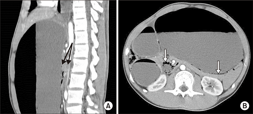
Abdominopelvic CT scans showed marked gastric distention and proximal duodenal dilatation with compression of the third segment between SMA and abdominal aorta. The SMA angle to the aorta is normally 45° (range, 38° to 56°), whereas in SMA syndrome, the SMA angle is decreased to 6° to 25° [8]. The patient’s aortomesenteric angle was decreased to 12°. Also, there were thin linear mural airs in the walls of the stomach and duodenum (Fig. 1). These findings were consistent with SMA syndrome. Based on the CT scans and his clinical symptoms, the patient was diagnosed as having gastric emphysema related with SMA syndrome. For gastric decompression, nasogastric tube was inserted and intravenous hydration was promptly started. Total parenteral nutrition was started from the day of admission, and he was observed closely in the intensive care unit. The intravenous broad spectrum antibiotics was added and to rule out the emphysematous gastritis, blood and gastric fluid were cultured.
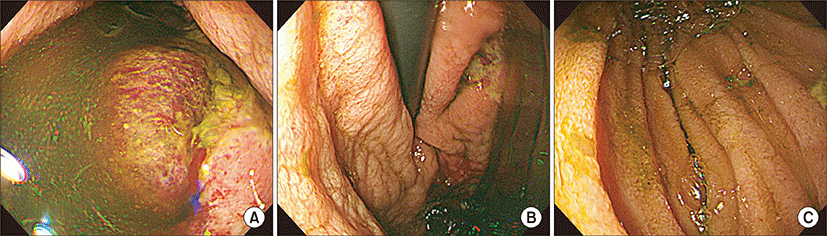
On the patient’s third day in hospital, upper gastrointestinal tract endoscopy revealed many geographic ulcers with mucosal hemorrhages in the body of the stomach. There was marked dilatation of the second segment of the duodenum with many geographic ulcers and a collapsed third segment associated with the SMA syndrome (Fig. 2). An intravenous proton pump inhibitor was administered to treat the peptic ulcer bleeding.
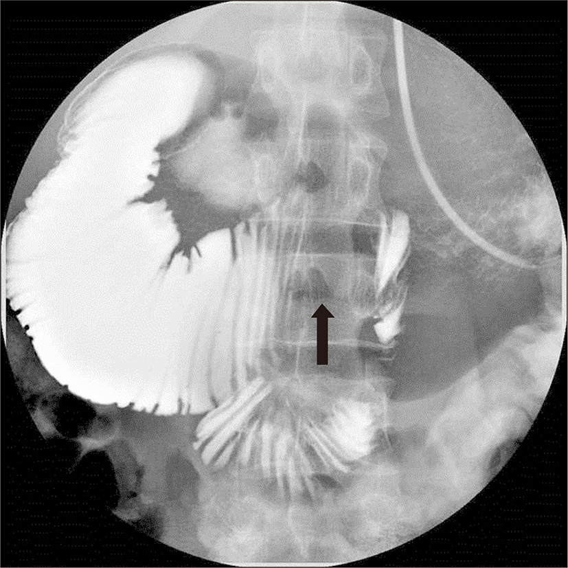
On the fourth day in hospital, a nasojejunal feeding tube was inserted with fluoroscopic guidance (Fig. 3). Nutritional support was performed with continuous enteral feeding. On the sixth day, results from a gastric fluid culture showed the presence of Enterococcus faecalis, but blood culture results were negative. On the ninth day in hospital, upper gastrointestinal series revealed a dilated proximal duodenum with a collapsed third portion. During this procedure, a small amount of contrast medium passed through from duodenum to jejunum over about 1 min. Based on these results, we decided to remove nasojejunal feeding tube and start oral feeding on the 9th hospital day. The patient was discharged and administered oral proton pump inhibitors 13 days after admission.
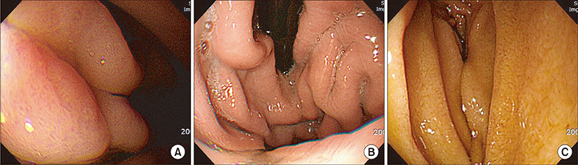
The patient improved without recurrence during a 3-month follow-up period. Follow-up upper endoscopy after 2 months showed complete recovery from the gastric emphysema and a mildly constricted third segment of the duodenum by extrinsic SMA compression (Fig. 4).
Discussion
Gastric emphysema is caused by increased intragastric pressure. Six cases have been reported in Korea since 2005 (Table 1) [9-14].Various causes of gastric emphysema have been reported: for example, severe vomiting [9], mucosal damage by methyl ethyl ketone peroxide ingestion [10], endoscopic submucosal dissection [11], endoscopic submucosal tumor biopsy [12], SMA syndrome due to anorexia nervosa [13], and SMA syndrome [14].
On the other hand, emphysematous gastritis is caused by infections with gas-forming microbes. Enterobacter species, Pseudomonas aeruginosa, Candida albicans, and Staphylococcus aureus are known as common causative organisms [15]. Emphysematous gastritis is associated with predisposing factors including ingestion of corrosive substances, alcohol abuse, abdominal surgery, diabetes, and immunosuppression [6]. In our case, the patient’s predisposing factors were anorexia nervosa with SMA syndrome, which induced duodenal obstruction and marked gastric distension. Therefore, his condition was a result of mucosal disruption of the stomach caused by increased intragastric pressure.
E. faecalis was cultured from the patient’s gastric fluid on the fourth day in hospital. It was difficult to differentiate his condition from emphysematous gastritis because of this infection. However, based on clinical symptoms and radiographic findings, the patient was diagnosed as having gastric emphysema.
The characteristic radiographic features of gastric emphysema are thin, linear streaks of air along the border of the stomach [16]. CT scans help to confirm this linear distribution of intramural air [17]. However, unlike gastric emphysema, emphysematous gastritis can demonstrate mottled mucosal fold thickening with irregular collections of gas within the stomach wall [17]. In our case, abdominopelvic CT scans showed thin linear air accumulations in the walls of the stomach and duodenum. They also revealed marked gastric distention and proximal duodenal dilatation with compression of the third segment between the SMA and abdominal aorta.
On early endoscopy, bubbles with a ‘cobblestone’ appearance can be seen in the gastric mucosa of patients affected with gastric emphysema or emphysematous gastritis [18]. Endoscopic findings in such patients include submucosal gas bubbles, necroinflammatory changes, and erosions [19]. In some cases, the mucosa appears normal [19]. In our case, in order to avoid barotrauma to the stomach, upper endoscopy was not done immediately. On the third day of hospital, when the patient’s physical condition became stable, upper endoscopy was performed very carefully. This demonstrated many gastric ulcers with mucosal hemorrhage in the body of the stomach. There were no mucosal bubbles or necrotic changes.
We have reported one similar case of gastric emphysema associated with SMA syndrome previously [14]. A 68-year-old man was diagnosed as having gastric emphysema that might have resulted from diabetic gastroparesis and SMA syndrome, associated with malnutrition. Emergent upper endoscopy revealed diffuse erosive and hemorrhagic lesions with clots from the distal esophagus to the lesser curvature of the stomach. No mucosal bubbles or necrotic changes were observed in that case either.
In the present case, the patient presented with abdominal distension, pain, and vomiting, as a result of increased intragastric pressure by duodenal obstruction. His rapid recovery with a benign clinical course confirmed our diagnosis.