Case Report

당뇨병 환자에서 발생한 피부 털곰팡이증

박지환, 박서화, 강은규, 경규천, 안효동, 안소연
Author Information & Copyright
홍익병원 내과
Department of Internal Medicine, Hongik Hospital, Seoul, Korea
Corresponding author So-Yeon An Department of Internal Medicine, Hongik Hospital, 225, Mokdong-ro, Yangcheon-gu, Seoul 07937, Korea Tel: 82-2-2600-0437, Fax: 82-2-2697-4605 E-mail: yeonigge00@naver.com

Copyright © 2016 Ewha Womans University School of Medicine. This is an open-access article distributed under the terms of the Creative Commons Attribution Non-Commercial License (http://creativecommons.org/licenses/by-nc/4.0) which permits unrestricted non-commercial use, distribution, and reproduction in any medium, provided the original work is properly cited.

Received: Sep 23, 2015; Accepted: Oct 29, 2015

Published Online: Jan 31, 2016

Abstract

Mucormycosis is a rare disease caused by fungi. Most commonly involved sites of mucormycosis infection are sinuses, lungs, skin and soft tissues. Systemic risk factors for mucormycosis are diabetes mellitus, neutropenia, corticosteroid use, hematological malignancies, organ transplantation, metabolic acidosis, deferoxamine use and advanced age. Local risk factors are history of trauma, burns, surgery and motor vehicle accidents. We present a case of cutaneous mucormycosis in a patient with diabetes mellitus. A 66-year-old female with uncontrolled diabetes mellitus, admitted with necrotizing lesion after minor abrasions on leg. We took a culture of the lesion and it is diagnosed with mucormycosis. Disease progressed despite administration of systemic amphotericin B. We performed above-knee amputation and changed antifungal agents into liposomal amphotericin B. A tissue biopsy showed nonseptate, irregularly wide fungal hyphae with frequent right-angle branching. Our case report suggests that patients with risk factors should be observed carefully.

Keywords: Mucormycosis; Diabetes mellitus; Trauma; Amputation; Liposomal amphotericin B

서 론

털곰팡이증은 드물지만 높은 사망률을 보이는 감염증으로, 접합균문 털곰팡이목에 속하는 진균에 의해 발생한다[1]. 털곰팡이증의 위험 인자로는 당뇨병, 호중구 감소증, 스테로이드 사용, 혈액학적 악성종양(백혈병, 림프종, 다발성 골수종), 재생불량성 빈혈, 골수형성이상증후군, 장기이식, 대사성 산증, deferoxamine 사용, 화상, 중증 외상 등이 있다[2]. 최근 전세계적으로 털곰팡이증의 발생이 증가하고 있는데, 특히 당뇨병 환자나 악성종양 환자에서 그 증가가 뚜렷하게 나타난다[1,3].

털곰팡이증은 비-안-대뇌형과 폐형, 위장관형, 피부형, 파종형 등의 다양한 형태로 나타나며 그 중 피부 털곰팡이증은 전체의 15%–20% 정도를 차지한다[4]. 피부 털곰팡이증은 주로 수술이나 화상, 토양에 오염되는 부상, 교통사고 등으로 인해 피부의 통합성이 깨지게 될 때 발생하게 된다[4].

이번 증례는 조절되지 않은 당뇨병이 있는 66세 환자로, 환자는 우측 다리에 가벼운 찰과상을 입은 후 발생한 심부 조직을 침범한 피부 털곰팡이증을 하지절단술 및 항진균제로 치료 후 완치 되었다. 피부 털곰팡이증의 증례는 국내에 보고된 바가 드물어 문헌고찰과 함께 보고하는 바이다.

증 례

환자는 66세 여자로 내원 10일 전 낙상하면서 발생한 우측 정강이 부위 찰과상으로 인한 우측 경골부위 통증 및 발열감을 주소로 인근병원에서 항생제 치료를 받았다. 수일 간의 치료에도 증상 호전 없이 괴사성 병변이 진행되어 적절한 치료 위해 본원 정형외과로 전원되었다. 10년 전 당뇨병을 진단받았으나 최근 5년간 특별한 치료를 받지 않은 과거력이 있으며, 흡연력 및 음주력, 가족력에서는 특이사항이 없었다.

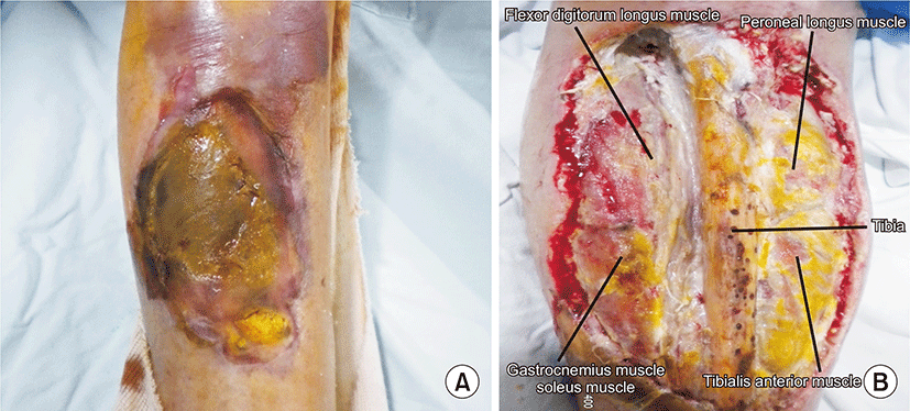
내원 당시 활력징후는 혈압 130/80 mmHg, 맥박 분당 72회, 체온 36.5°C로 정상이었고, 환자 신장은 147 cm, 체중은 35 kg이었다. 이학적 검사에서 우측 정강이 앞쪽 부위로 경골이 노출되어 있었고 피부와 전경골근의 농양 및 경골의 괴사가 동반되어 있었다(Fig. 1A). 말초혈액검사에서 백혈구 28,200/μL (중성구 비율 92%)으로 증가되어 있었고, 혈색소 13.5 g/dL, 혈소판 277,000/μL이었다. 생화학적 검사에서 적혈구침강속도 30 mm/hr, C반응성단백 18.61 mg/dL으로 증가되어 있었고, 혈액요소질소 15.8 mg/dL, 크레아티닌 0.5 mg/dL 및 나트륨, 칼륨, 요산, 칼슘, 인은 정상범위에 있었다. 당화 혈색소는 13.7 %로 증가되어 있었다. 내원하여 촬영한 하지 전산화 단층촬영 사진에서는 경골 돌출 소견 이외에는 특이 소견 보이지 않았다.

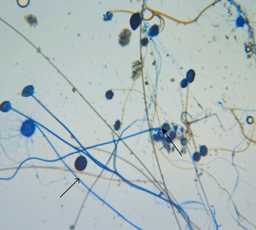
내원 당일 시행한 농양의 배양검사 결과가 나오기 전까지 경험적 항생제 tazobactam/piperacillin (Tazoperan, Chong Kun Dang Pharmaceutical Corp., Seoul, Korea) 2.25 g과 levofloxacin (Cravit, Jeil Ltd., Seoul, Korea) 750 mg을 정맥 투여하였고, 입원 5일째 농양의 배양검사 결과 Rhizopus Spp.가 확인되어(Fig. 2) amphotericin B 35 mg (1 mg/kg)을 24시간 마다 정주로 투여하였다. 조절되지 않는 당뇨병에 대해서는 insulin detemir (Levemir, Novo Nordisk Pharma Korea, Seoul, Korea), insulin aspart (NovoRapid, Novo Nordisk Pharma Korea, Seoul, Korea)로 조절하였다.

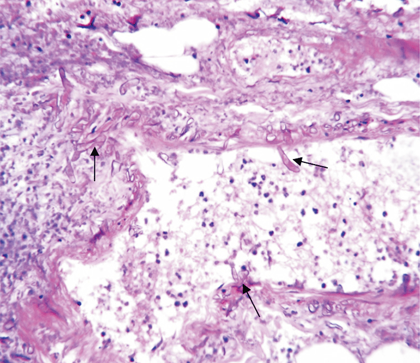
지속적인 변연 절제술과 항진균제 투여에도 불구하고 피부 털곰팡이증이 근육과 뼈까지 침범하는 양상(Fig. 1B)이었기 때문에 더 이상의 변연 절제가 불가능하다고 생각되어, 입원 21일째에 우측 슬관절 상부 절단술을 시행하였으며 수술 소견에서 복재 정맥, 림프 조직까지 털곰팡이증의 침범이 확인되었다. 절단된 다리의 단면 조직의 현미경 소견에서 혈관벽을 침범한 직각으로 분지하는 넓고, 불규칙적인 모양의 무격균사가 관찰 되었다(Fig. 3). 환자 지속적인 발열 소견 보이고 전신 상태 악화되어 입원 26일째 본원 내과로 전과되었다. 수술 이후 혈액요소질소 및 크레아티닌 검사 결과 지속적 상승 보여(혈액요소질소 54.8 mg/dL, 크레아티닌 2.8 mg/dL) 항진균제를 liposomal amphotericin B 150 mg으로 변경하여 24시간 마다 정주 투여하였다. Liposomal amphotericin B로 변경 투여 후 신장기능 검사 및 백혈구 증가증의 호전 및 발열 등의 임상 양상 호전 되어 입원 45일까지 사용하였고, 입원 61일째 요양병원으로 전원하였다.

고 찰

피부 털곰팡이증은 전체 털곰팡이증의 15%–20%정도를 차지하며 당뇨병환자에서는 4번째로 흔하게 보이는 형태이다. 이중 관통상과 연관된 것이 34%, 수술 또는 드레싱과 연관된 것이 각각 15%, 화상이 6% 정도의 분포를 보이고 있다[4,5]. 피부 털곰팡이증은 20종류 이상의 Mucorales 종에 의해 발생하며 그 중 토양에서 흔히 발견되는 Mucor, Rhizopus가 가장 흔한 원인 균종이다[6].

피부 병변은 다양한 정도의 중심부위 괴사를 동반한 통증, 홍반, 경결을 특징으로 하고 좀 더 진행된 경우 괴저와 괴사성 근막염의 형태를 보인다[7]. 하지만 백선이나 다형 홍반 같은 형태 등 다양한 피부 병변을 보이는 경우도 있다[6]. 피부 털곰팡이증은 감염의 침범 범위에 따라 외피와 피하조직에 국한된 형태, 뼈, 인대, 근육층과 같은 심부 조직을 침범한 형태, 인접하지 않은 다른 장기로 파종된 형태로 나뉘며[8], 대부분 외피에 국한되어 발생하나, 24% 정도에서는 뼈, 인대, 근육층과 같은 심부 조직까지 침범하고, 피부에서 인접하지 않은 다른 장기로의 혈행성 파종은 20%, 다른 장기에서 피부로 혈행성 파종이 되는 경우는 3% 정도에서 발생한다[5].

털곰팡이증은 다양한 임상 양상을 보이기 때문에 조기 진단이 어렵다. 하지만 위험 인자를 가지고 있는 환자거나 경험적 항생제에 반응 없이 악화되는 경과를 보이는 환자의 경우 털곰팡이증의 가능성을 염두에 두고 검사를 진행해야 한다. 털곰팡이증은 혈청학적 검사 및 영상 검사로 진단하기 어려우며 조직병리학적 검사 및 배양 검사가 필수적이다. 조직병리학적으로, 직각으로 분지하는 10–50 μm의 넓고, 불규칙적인 모양의 무격균사가 보이고 혈전에 의한 혈관 침범과 조직 괴사가 보일 때 진단할 수 있다[3]. 배양 검사에서 양성이 나오는 경우는 50% 정도이며 Rhizopus spp.는 가장 많이 동정되는 균 중 하나이다[2]. 최근에는 중합효소 연쇄반응 검사를 통해 빠른 진단이 이루어진 증례들이 보고 되었으나 이 진단 기법에 대해서는 좀 더 연구가 필요하다.

피부 털곰팡이증은 Pseudomonas, Aspergillus, Histoplasma 등에 의한 연조직 감염, 혈관염, 괴저고름피부증 등과 임상적 양상만으로 구별이 힘들다. 감별 진단을 위해 초기에 빠른 조직병리학적 검사 및 배양 검사를 시행하여 특이적인 곰팡이의 확인이 필요하며 보조적으로 Hematoxylin & Eosin 염색, Periodic acid-Schiff 염색, Gomori’s Methenamine Silver 염색이 필요하다. 또한 여러 부위에서의 조직 검사 및 반복 검사가 도움이 된다[9-11].

본 증례의 경우 발병 초기에 배양 검사와 조직병리학적 검사를 시행하였다면, 다른 질환과의 감별 진단 후 피부 털곰팡이증에 대한 빠르고 정확한 치료를 시행할 수 있었을 것으로 생각 된다. 본 증례에서는 내원 당시 절개 및 배농 부위 배양 검사에서 Rhizopus spp.를 확인하였고 수술 후 다리의 절단된 단면 조직검사에서 혈관을 침범한 무격균사가 확인되어서 털곰팡이증으로 진단하고 이에 따른 치료를 시행하였다.

털곰팡이증의 치료원칙은 조기 진단과 위험 인자의 제거, 감염조직의 적절한 외과적 절제, 효과적인 항진균제 요법이다[12,13]. 털곰팡이증은 혈전에 의한 혈관 침범과 조직 괴사라는 특징 때문에 항진균제가 효과적으로 염증부위에 도달하지 못하므로 감염원을 제거하기 위한 적극적인 외과적 절제와 전신적 항진균제의 사용이 필수적이다[8]. 항진균제로는 liposomal amphotericin B, 5–10 mg/kg/day가 1차 치료제이며 amphotericin B deoxycholate 제재는 신독성으로 더 이상 사용이 권고되지 않는다. Liposomal amphotericin B를 이용한 치료에 실패하거나 치명적인 약제 부작용이 발생하였을 때는 posaconazole을 사용하거나 liposomal amphotericin B와 posaconazole을 함께 사용하는 것을 권고하고 있다[14]. 치료 기간에 대해서는 명확하게 정해진 바 없이 환자 상태에 따라 치료하게 되는데[15], 본 증례에서는 침범한 다리의 절단 후 국소적, 전신적으로 염증의 증거가 보이지 않고 임상적으로 안정화 될 때까지 충분히 항진균제 사용 후 중단하였다.

피부 털곰팡이증은 다른 형태의 털곰팡이증보다는 좋은 예후를 보이지만, 여전히 높은 사망률을 보이는 중대한 감염질환으로, 침범 범위에 따라 4%–94%까지 다양한 사망률을 보인다[5]. 또한, 진단이 늦어지거나, 항진균제 치료나 위험 인자의 교정이 적절하게 이루어 지지 못할 경우 사망률은 더 증가한다[2,15].

이에 저자들은 혈당이 조절되지 않는 66세 환자에서 발생한 심부 조직을 침범한 피부 털곰팡이증을 하지절단술 및 전신적인 항진균제로 치료한 증례를 통해 위험 인자를 가진 환자에서의 적절한 의심과 빠른 진단 및 치료의 중요성을 환기하고자 한다.

REFERENCES

1.

Camara-Lemarroy CR, Gonzalez-Moreno EI, Rodriguez-Gutierrez R, Rendon-Ramirez EJ, Ayala-Cortes AS, Fraga-Hernandez ML, et al. 2014; Clinical features and outcome of mucormycosis. Interdiscip Perspect Infect Dis. 2014:562610

2.

Mantadakis E, Samonis G. 2009; Clinical presentation of zygomycosis. Clin Microbiol Infect. 15(Suppl 5):15-20

3.

Ingram PR, Suthananthan AE, Rajan R, Pryce TM, Sieunarine K, Gardam DJ, et al. 2014; Cutaneous mucormycosis and motor vehicle accidents: findings from an Australian case series. Med Mycol. 52:819-825

4.

Zaman K, Kaur H, Rudramurthy SM, Singh M, Parashar A, Chakrabarti A. 2015; Cutaneous mucormycosis of scalp and eyelids in a child with type I diabetes mellitus. Indian J Dermatol Venereol Leprol. 81:275-278

5.

Roden MM, Zaoutis TE, Buchanan WL, Knudsen TA, Sarkisova TA, Schaufele RL, et al. 2005; Epidemiology and outcome of zygomycosis: a review of 929 reported cases. Clin Infect Dis. 41:634-653

6.

Petrikkos G, Skiada A, Lortholary O, Roilides E, Walsh TJ, Kontoyiannis DP. 2012; Epidemiology and clinical manifestations of mucormycosis. Clin Infect Dis. 54(Suppl 1):S23-S34

7.

Ribes JA, Vanover-Sams CL, Baker DJ. 2000; Zygomycetes in human disease. Clin Microbiol Rev. 13:236-301

8.

Iida T, Sawada N, Takahashi M, Zendejas IR, Kayler LK, Magliocca JF, et al. 2010; Successful treatment of invasive mucormycosis in a liver transplant patient by arm amputation. Transplant Proc. 42:2794-2796

9.

Su WP, Davis MD, Weenig RH, Powell FC, Perry HO. 2004; Pyoderma gangrenosum: clinicopathologic correlation and proposed diagnostic criteria. Int J Dermatol. 43:790-800

10.

Kerr OA, Bong C, Wallis C, Tidman MJ. 2004; Primary cutaneous mucormycosis masquerading as pyoderma gangrenosum. Br J Dermatol. 150:1212-1213

11.

Geller JD, Peters MS, Su WP. 1993; Cutaneous mucormycosis resembling superficial granulomatous pyoderma in an immunocompetent host. J Am Acad Dermatol. 29:462-465

12.

Luo Y, Zeng F, Huang X, Li Q, Tan G, Xi L, et al. 2014; Successful treatment of a necrotizing fasciitis patient caused by Mucor indicus with amphotericin B and skin grafting. Mycopathologia. 177:187-192

13.

de Oliveira-Neto MP, Da Silva M, Fialho Monteiro PC, Lazera M, de Almeida Paes R, Novellino AB, et al. 2006; Cutaneous mucormycosis in a young, immunocompetent girl. Med Mycol. 44:567-570

14.

Cornely OA, Arikan-Akdagli S, Dannaoui E, Groll AH, Lagrou K, Chakrabarti A, et al. 2014; ESCMID and ECMM joint clinical guidelines for the diagnosis and management of mucormycosis 2013. Clin Microbiol Infect. 20(Suppl 3):5-26

15.

Ahmadinejad Z, Khazraiyan H, Ghanbari F, Ahmadi B, Gerami Shoar M. 2013; Cutaneous Mucormycosis in a Diabetic Patient following Traditional Dressing. Case Rep Dermatol Med. 2013:894927
emj-39-010-f1
Fig. 1. (A) A photograph of the right shin. The tibia is exposed on the front of the right shin accompanying necrotizing tibialis anterior muscle. (B) The picture of the wound during debridement before the surgery. Portions of tibia, tibialis anterior muscle and gastrocnemius muscle are seen. Portions of peroneal longus muscle and soleus muscle are demonstrated, as well.
Download Original Figure
emj-39-010-f2
Fig. 2. Rhizopus spp. was found and identified in the experiment cultivating Rhizopus hyphae in pus with debridement which cultivated no septum. It formed a root-shaped hyphae (rhizoid; closed arrow) and an unbranched sporangiospore shaped object (open arrow) at the joining part between the stolon and sporangiospore.
Download Original Figure
emj-39-010-f3
Fig. 3. Many zygomycetes (nonseptate, irregularly wide fungal hyphae with frequent right-angle branching; closed arrow) invaded the blood vessel wall (H&E, ×400).
Download Original Figure