Case Report

An Esophageal Leiomyoma with Cystic Degeneration Mimicking a Malignant Neoplasm

Gwang Ha Kim1,2,*https://orcid.org/0000-0001-9721-5734, Dong Chan Joo1https://orcid.org/0000-0001-8734-4938, Moon Won Lee1https://orcid.org/0000-0002-8411-6398, Bong Eun Lee1https://orcid.org/0000-0003-2734-2134, Kyungbin Kim3https://orcid.org/0000-0001-5430-4235
Author Information & Copyright
1Department of Internal Medicine, Pusan National University School of Medicine, Busan, Korea
2Biomedical Research Institute, Pusan National University Hospital, Busan, Korea
3Department of Pathology, Pusan National University Hospital, Busan, Korea
*Corresponding author: Gwang Ha Kim, Department of Internal Medicine, Pusan National University School of Medicine, 179 Gudeok-ro, Seo-gu, Busan 49241, Korea, Tel: 82-51-240-7869, Fax: 82-51-244-8180, E-mail: doc0224@pusan.ac.kr

© Copyright 2023 Ewha Womans University College of Medicine and Ewha Medical Research Institute. This is an Open-Access article distributed under the terms of the Creative Commons Attribution Non-Commercial License (http://creativecommons.org/licenses/by-nc/4.0/) which permits unrestricted non-commercial use, distribution, and reproduction in any medium, provided the original work is properly cited.

Received: Sep 06, 2023; Accepted: Oct 06, 2023

Published Online: Oct 31, 2023

ABSTRACT

Esophageal subepithelial tumors (SETs) are commonly encountered during screening endoscopy, and leiomyomas are the most common SET of the esophagus. Almost all patients with esophageal leiomyomas are asymptomatic; however, some present with dysphagia, depending on the size of the tumor and the extent to which it encroaches on the lumen. The typical endosonographic features of esophageal leiomyomas include well-demarcated, homogeneously hypoechoic lesions with echogenicity similar to that of the surrounding proper muscle layer, but without cystic changes. Histopathologically, esophageal leiomyomas do not undergo cystic or myxoid degeneration. This report presents a case involving a 65-year-old man with a symptomatic esophageal SET and endosonographic features indicative of malignant neoplasms, who was diagnosed with esophageal leiomyoma with cystic and myxoid degeneration following surgical resection.

Keywords: Endoscopic ultrasound; Esophagus; Leiomyoma; Mesenchymal tumors

Introduction

Esophageal subepithelial tumors (SETs) are commonly encountered during screening endoscopy, with an incidence of 0.4% (246/65,233), based on a recent Korean multicenter study [1]. Almost all esophageal SETs are asymptomatic and clinically insignificant, and leiomyomas are the most common. However, malignant SETs, such as gastrointestinal stromal tumors (GISTs) and SET-like carcinomas, can also occur in the esophagus. Endoscopic ultrasonography (EUS) is the most useful diagnostic modality for evaluating SETs of the gastrointestinal tract. The typical EUS features of leiomyomas include well-demarcated, homogeneously hypoechoic lesions with echogenicity similar to that of the surrounding proper muscle layer [24]. Calcifications are relatively common in leiomyomas (6.5%–18%); however, cystic changes are rarely observable [5,6]. In contrast, GISTs exhibit typical features on EUS, such as heterogeneously hypoechoic lesions with a marginal halo and hyperechoic spots, and slightly increased echogenicity compared to the surrounding proper muscle layer [5]. Furthermore, irregular margins and cystic changes have been suggested to be predictive factors of malignant potential [7]. Herein, we report a case of symptomatic esophageal leiomyoma with cystic changes on EUS that was managed successfully using thoracoscopic resection.

Case

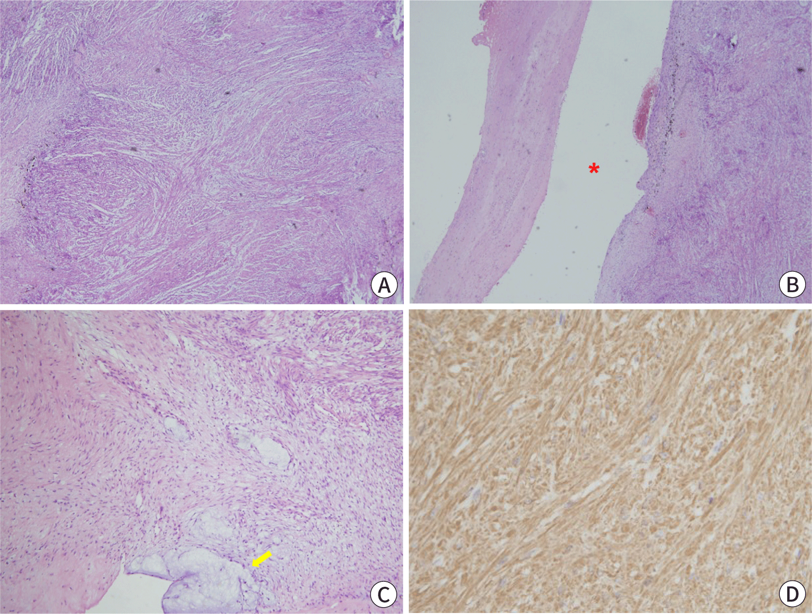
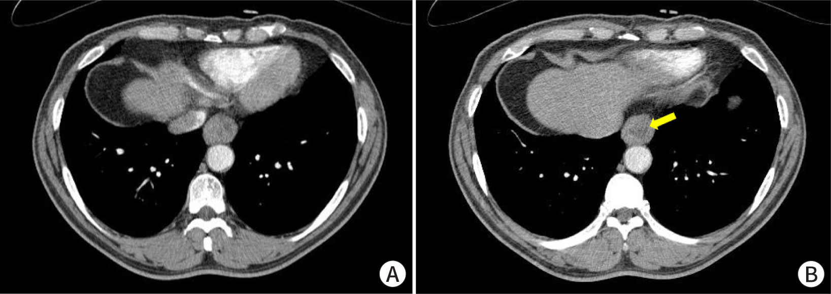
A 65-year-old man presented with an esophageal SET that was incidentally detected during esophagogastroduodenoscopy. The patient had a 1-year history of chest discomfort and dyspepsia, and a 5-year history of diabetes. On endoscopy, the tumor was observed in the lower esophagus and no erosion or ulceration was noted on its surface (Fig. 1A). The tumor was hard and immovable when pressed with biopsy forceps. On EUS, the tumor measured 3.0 cm and had a heterogeneously hypoechoic appearance in the proper muscle layer (Fig. 1B). A cystic change, measuring 1.5 cm, was also noted on the inner side of the tumor (Fig. 1C). These EUS features are suggestive of malignant neoplasms such as GISTs. Physical examination revealed no abnormalities. Laboratory investigations revealed the following: white blood cell count, 5,470 cells/μL; hemoglobin, 15.0 g/dL; platelet count, 193,000 cells/μL; prothrombin time, 10.9 s (international normalized ratio 0.98); aspartate aminotransferase, 32 IU/L; alanine aminotransferase, 45 IU/L; total bilirubin, 0.62 mg/dL; total protein, 7.9 g/dL; albumin, 4.6 g/dL; blood urea nitrogen, 14.3 mg/dL; and creatinine, 0.9 mg/dL. Electrocardiography results were normal. Contrast-enhanced chest CT revealed asymmetric wall thickening with heterogeneous enhancement and a non-enhanced area in the lower esophagus, suggestive of malignant lesions (Fig. 2). Because the patient experienced symptoms, including chest discomfort, and EUS and CT findings were highly suspicious for malignant neoplasms, thoracoscopic resection was performed. Macroscopic examination revealed a well-defined, solid white ovoid mass with cystic changes, measuring 3.2 cm×2.2 cm, in the muscularis propria of the esophagus. Histopathological examination of the specimen revealed intersecting bundles or fascicles of bland spindle cells with eosinophilic cytoplasm (Fig. 3A). Cystic and myxoid degeneration was observed; however, significant cytological atypia, increased mitotic activity, and tumor necrosis were not detected (Fig. 3B, C). Immunohistochemical examination revealed that the tumor cells were positive for smooth muscle actin (Fig. 3D) and desmin, negative for c-kit, CD34, and S-100, and had a low Ki-67 proliferation index (<1%). Accordingly, the tumor was diagnosed as leiomyoma with cystic and myxoid degeneration. The patient’s symptoms, including chest discomfort, subsided completely, and no recurrence was observed at the 24-month follow-up.

emj-46-4-19-g1
Fig. 1. Endoscopic and endosonographic findings. (A) A subepithelial tumor, measuring 3 cm, without surface erosion or ulceration is evident in the lower esophagus. (B, C) Endoscopic ultrasonography reveals a heterogeneously hypoechoic lesion with cystic changes (arrow) in the proper muscle layer.
Download Original Figure
emj-46-4-19-g2
Fig. 2. Contrast-enhanced chest CT. (A, B) Asymmetric wall thickening with heterogeneous enhancement and a non-enhanced area (arrow) is evident in the lower esophagus.
Download Original Figure
emj-46-4-19-g3
Fig. 3. Histopathological findings. (A) Intersecting bundles or fascicles of bland spindle cells with eosinophilic cytoplasm are observed (hematoxylin and eosin stain, ×40). (B, C) Cystic (asterisk) and myxoid degeneration (arrow) is evident; however, significant cytological atypia, increased mitotic activity, and tumor necrosis are not detected (hematoxylin and eosin stain, ×40 and ×100, respectively). (D) Tumor cells are positive for smooth muscle actin stain (×400).
Download Original Figure

Discussion

Esophageal leiomyomas are the most common benign SETs in the esophagus [8]. Unlike GISTs, esophageal leiomyomas do not undergo cystic or myxoid degeneration and are very rarely ulcerated [9]. Almost all patients with esophageal leiomyomas are asymptomatic, but some present with dysphagia, depending on the size of the tumor. In this case, the tumor was not ulcerated, but did exhibit cystic degeneration. This was believed to be the cause of the patient’s symptoms (chest discomfort), which subsided after thoracoscopic resection.

Virtually all esophageal leiomyomas are well-demarcated, homogeneously hypoechoic lesions in the muscularis mucosa or proper muscle layer on EUS, and cystic changes are rarely observed [2,5]. However, in the present case, the esophageal leiomyoma appeared as a well-demarcated but heterogeneously hypoechoic lesion in the proper muscle layer. In addition, the echogenicity of the tumor was greater than that of the surrounding proper muscle layer, and extensive cystic changes were observed. These EUS features are usually highly suggestive of malignant stromal tumors such as GISTs. Chest CT also suggested a malignant rather than a benign lesion. However, EUS and CT cannot perfectly differentiate between benign and malignant SETs. Therefore, EUS-guided fine-needle biopsy has recently emerged as a commonly used diagnostic modality, with an acceptable yield (87.9%) [6,10,11]. In the present case, because the SET was symptomatic and the EUS and CT features were highly suggestive of malignancy, the tumor was surgically resected without histopathological confirmation via EUS-guided fine-needle biopsy.

Histopathologically, both leiomyomas and GISTs are mainly composed of spindle cells and display smooth muscle or nerve sheath differentiation; as such, immunohistochemical analysis is necessary for an accurate diagnosis. Leiomyomas are defined as desmin-positive and c-kit-negative tumors, whereas GISTs are defined as c-kit-positive [12]. The patient described in the present case was ultimately diagnosed with leiomyoma based on immunohistochemical staining.

Because esophageal leiomyomas are benign, resection is not required in most cases. However, when patients experience symptoms, such as dysphagia, the tumor size increases on follow-up examinations or, if the patient strongly desires to undergo tumor resection, resection is needed. In the past, surgical resection, such as thoracoscopic resection, was the primary treatment modality for esophageal leiomyomas located in the muscularis propria. However, endoscopic resection techniques, such as endoscopic submucosal dissection, endoscopic submucosal excavation, and submucosal tunneling endoscopic resection, have recently been introduced and have yielded high complete resection rates [13]. However, when a malignant SET is highly suspected, as in the present case, surgical resection is necessary to achieve satisfactory oncological outcomes.

Esophageal leiomyomas are often encountered as benign lesions during endoscopy. However, these tumors are sometimes symptomatic and do not exhibit the typical EUS and CT features of leiomyomas. In this situation, surgical resection is required to alleviate symptoms and rule out the possibility of malignancy.

Acknowledgements

This case was previously published in Korean J Helicobacter Up Gastrointest Res 2021;21:152-155 in Korean. However, this case report includes different endoscopic, radiologic, and histopathologic images and is written in English to present different aspects. It is submitted with the approval of the editor of the Korean J Helicobacter Up Gastrointest Res.

Conflict of Interest

No potential conflict of interest relevant to this article was reported.

Author Contribution

Conceptualization: Kim GH

Formal Analysis: Joo DC, Lee MW

Investigation: Lee BE, Kim K

Methodology: Kim GH, Kim K

Project Administration: Kim GH, Kim K

Writing – Original Draft: Kim GH

Writing – Review & Editing: Kim GH, Joo DC, Lee MW, Lee BE, Kim K

Ethics Approval and Consent to Participate

Not applicable.

References

1.

Choe Y, Cho YK, Kim GH, Choi JH, Kim ES, Kim JH, et al. Prevalence, natural progression, and clinical practices of upper gastrointestinal subepithelial lesions in Korea: a multicenter study. Clin Endosc. 2023 Aug; 25 [Epub]

2.

Lee MW, Kim GH. Diagnosing gastric mesenchymal tumors by digital endoscopic ultrasonography image analysis. Clin Endosc. 2021; 54((3)):324-328

3.

Kim DU, Kim GH, Ryu DY, Lee DG, Cheong JH, Lee BE, et al. Endosonographic features of esophageal granular cell tumors using a high-frequency catheter probe. Scand J Gastroenterol. 2011; 46((2)):142-147

4.

Seo WY, Kim GH. H I. An esophageal leiomyoma mistaken as an esophageal duplication cyst. Korean J Helicobacter Up Gastrointest Res. 2021; 21((2)):152-155

5.

Kim GH, Park DY, Kim S, Kim DH, Kim DH, Choi CW, et al. Is it possible to differentiate gastric GISTs from gastric leiomyomas by EUS?. World J Gastroenterol. 2009; 15((27)):3376-3381

6.

Lee JS, Cho CM, Kwon YH, Seo AN, Bae HI, Han MH. Comparison of diagnostic performances of slow-pull suction and standard suction in endoscopic ultrasound-guided fine needle biopsy for gastrointestinal subepithelial tumors. Clin Endosc. 2022; 55((5)):637-644

7.

Shah P, Gao F, Edmundowicz SA, Azar RR, Early DS. Predicting malignant potential of gastrointestinal stromal tumors using endoscopic ultrasound. Dig Dis Sci. 2009; 54((6)):1265-1269

8.

Ryu DG, Choi CW. Common gastric subepithelial tumors in Koreans. Korean J Helicobacter Up Gastrointest Res. 2022; 22((1)):29-37

9.

Levine MS. Benign tumors of the esophagus.In In: Gore RM, Levine MS, editors.editors Textbook of gastrointestinal radiology. 2nd ed Philadelphia: W.B. Saunders. 2000; p p. 387-402.

10.

Facciorusso A, Sunny SP, Del Prete V, Antonino M, Muscatiello N. Comparison between fine-needle biopsy and fine-needle aspiration for EUS-guided sampling of subepithelial lesions: a meta-analysis. Gastrointest Endosc. 2020; 91((1)):14-22

11.

Nakatani S, Okuwaki K, Watanabe M, Imaizumi H, Iwai T, Matsumoto T, et al. Stereomicroscopic on-site evaluation in endoscopic ultrasound-guided tissue acquisition of upper gastrointestinal subepithelial lesions. Clin Endosc. 2023 Apr; 18 [Epub]

12.

Fletcher CDM, Berman JJ, Corless C, Gorstein F, Lasota J, Longley BJ, et al. Diagnosis of gastrointestinal stromal tumors: a consensus approach. Hum Pathol. 2002; 33((5)):459-465

13.

Nabi Z, Reddy DN. Submucosal endoscopy: the present and future. Clin Endosc. 2023; 56((1)):23-37

PDF Download
PubReader
Export Citation
Email the Author

Metrics

View
877
Download
441
Crossref
0
An Esophageal Leiomyoma with Cystic Degeneration Mimicking a Malignant Neoplasm

QR Code of this Article:


Related Articles

A Case of Multiple Leiomyoma of the Scrotum
Ewha Med J 1993;16(2):205-208.

A Case of Leiomyoma of the Vulva
Ewha Med J 1994;17(2):173-176.

A Case of Leiomyoma in the Female Paraurethra
Ewha Med J 1995;18(2):149-151.

3 Cases of Leiomyoma in the Female Urethra
Ewha Med J 1991;14(2):191-194.

Leiomyoma Arising From Distal Phalanx of Thumb
Ewha Med J 2010;33(1):35-38.