
Introduction
Total mesorectal excision (TME) is established as the prevailing therapeutic approach for rectal cancer, facilitating comprehensive tumor eradication through excision of the primary tumor along with the enveloping mesorectum and associated regional lymph nodes. This technique has demonstrated commendable outcomes in terms of local disease control and prolonged survival rates. Nevertheless, notable morbidities have been documented, including anastomotic leakage, genitourinary impairment, and the necessity for temporary or permanent stoma formation [1-3].
There has been a growing interest in the exploration of local treatment options for rectal cancer due to the significant morbidities associated with TME. This interest stems from the possibility of curing some patients without resorting to radical surgery, which often comes with its own set of disadvantages [4,5]. Local excision (LE) is an appealing choice for early rectal cancer, as it can potentially spare patients from unnecessary extensive rectal resection and the related complications. This approach offers several potential advantages, including reduced postoperative pain, quicker recovery, and the preservation of anorectal function without the need for a stoma. Additionally, new techniques for LE, such as transanal endoscopic microsurgery (TEM) and transanal minimally invasive surgery (TAMIS), have been introduced to address the limitations of conventional transanal excision (TAE).
Notwithstanding its advantages, there exists a concern regarding the potential risk of lymph node metastasis associated with transanal LE, which may hinder its ability to deliver oncologic outcomes equivalent to those of TME [6]. Multiple studies have reported a higher incidence of local recurrence with LE than with TME in the treatment of early rectal cancer [7,8]. These observations underscore the pivotal role of meticulous patient selection in guiding the choice of appropriate treatment modalities.
This review article seeks to provide a comprehensive overview of the various surgical techniques utilized in transanal LE for early rectal cancer, delving into indications, patient selection criteria, and technical considerations, while also emphasizing both their advantages and limitations.
Patient Selection
Precise patient selection, coupled with the meticulous execution of full-thickness, margin-free excisions, has a major effect on patient outcomes following LE for rectal cancer. In appropriately chosen individuals, the incidence of local recurrence has been documented to fall below 4%, rendering LE a potentially curative treatment option that yields comparable oncological results to radical surgery [4]. Consequently, it is imperative for surgeons to endeavor to differentiate between patients who are at high or low risk for local recurrence and lymph node metastasis prior to considering LE as a therapeutic approach for rectal cancer.
To ensure the appropriate selection of patients who are likely to benefit the most from LE, it is imperative to commence with a digital rectal examination, which clarifies key parameters such as tumor mobility, the distance from the anal verge, and the condition of the anal sphincter. Subsequently, proctoscopy aids in the evaluation of more proximal tumors, providing valuable insights into their dimensions and proximity to the anal verge. LE is generally considered technically feasible if the tumor occupies a maximum of 30% of the bowel circumference, measures no more than 3 cm, and exhibits mobility.
Preoperative investigations in the evaluation of rectal cancer encompass a range of diagnostic modalities, including radiological, endoscopic, and histological approaches where feasible. Typically, a comprehensive assessment involves CT scans of the chest, abdomen, and pelvis, complemented by PET scans when equivocal CT findings are present, to determine the presence of distant metastasis. For the locoregional evaluation of rectal cancer, MRI and endo-rectal ultrasonography (ERUS) or their combination is employed. ERUS has been proposed to exhibit superior accuracy in early disease staging when compared to MRI, albeit with reduced precision in assessing lymph node involvement. Conversely, MRI demonstrates superior accuracy in assessing lymph node status compared to ERUS [9,10].
An endoscopic examination serves the purpose of localizing lesions from the anal verge, conducting biopsies for histological evaluation, and estimating the degree of submucosal invasion through the analysis of glandular crypt patterns. Historically, rectal lesions situated within 10 cm of the anal verge were deemed suitable candidates for LE due to the limitations of surgical access and suboptimal tumor visualization. However, advances in technology and instrumentation have made it possible to reach higher lesions with improved visualization using the endoscopic approach. Innovative techniques such as TEM and TAMIS have extended access to lesions located up to 15 cm within the rectum. During an endoscopic evaluation for a primary rectal lesion, the submucosal invasion depth can be estimated by evaluating glandular crypt patterns. The presence of regular pit patterns typically signifies lesions confined to the mucosal layer, which makes them amenable to endoscopic resection. Conversely, the identification of irregular pit patterns, characterized by architectural distortion and amorphous structures, suggests an elevated risk of deep submucosal invasion [11,12].
Several histological parameters have demonstrated predictive value in assessing the likelihood of invasive disease and the risk of lymph node metastasis after endoscopic biopsy or resection. A consensus exists that resection margins equal to or greater than 1 mm are generally deemed sufficient, while margins less than 1 mm have been associated with recurrence rates of up to 33% [13,14]. Of paramount importance among these risk factors is the depth of tumor infiltration, as the risk of lymph node metastases steadily escalates with increased submucosal infiltration in early rectal cancer. The subclassification of T1 cancers into three tiers of submucosal invasion has shown a correlation with lymph node metastatic risk: 0%−3% for sm1, 8%−11% for sm2, and 11%−25% for sm3 invading tumors [15]. In accordance with a comprehensive cohort study, T2 rectal cancers exhibit a 21% risk of lymph node metastasis [16]. Furthermore, other indicators of aggressive tumor behavior include suboptimal histological grade, mucinous tumors, signet ring cell tumors, and the presence of lymphovascular invasion or perineural invasion [17]. Lastly, the presence of isolated clusters of malignant cells at the leading edge of the tumor, referred to as tumor budding, has also demonstrated a significant association with unfavorable oncological outcomes [14]. Occasionally, these characteristics are only definitively identified through pathological specimen review following LE, making it necessary to consider additional treatment modalities.
In summary, the optimal candidate for a LE procedure is a rectal adenocarcinoma smaller than 3 cm, classified as T1 and limited to the sm1 layer, exhibiting well-differentiated characteristics, and devoid of tumor budding, lymphovascular invasion, or perineural invasion, with an exceedingly low likelihood of lymph node metastasis. Conversely, if preoperative assessments reveal the presence of high-risk features, careful consideration should be given to the appropriateness of pursuing LE, and it may be regarded as an indication for palliative management.
Surgical Techniques of Transanal Local Excision
Tumors located within 10 cm of the anal verge can be surgically resected using conventional TAE. This procedure necessitates prior bowel preparation and the administration of prophylactic antibiotics. The patient's positioning during surgery is determined by the tumor's specific location; posterior tumors require lithotomy positioning, while anterior and lateral tumors are excised with the patient in the prone jackknife position. Anesthesia options encompass both general and regional techniques. To facilitate exposure, anal dilation is achieved using instruments like a Parks retractor or lone-star retractor, with the potential addition of lateral traction sutures to enhance visibility. Electrocautery is employed to create a radial line of dissection, ensuring a 1 cm margin. The rectal excision is performed as a full-thickness procedure, reaching the mesorectal fat. The closure of the rectal wall defect is transverse to prevent luminal narrowing, employing either a continuous or interrupted absorbable suture. Finally, the specimen is securely affixed to a board to facilitate a precise pathological assessment of the oriented margins.
In the 1980s, Buess et al. introduced TEM, employing a 4-cm-diameter rigid rectoscope equipped with a magnified binocular viewer, which facilitated a three-dimensional stereoscopic visualization of the rectum [18]. This instrument was inserted into the anus, creating an airtight seal to permit rectal insufflation using CO2 at pressures ranging from 10 to 15 mmHg, achievable through conventional laparoscopic CO2 insufflators [5,19]. The magnified view enabled the examination of approximately 220° s of the rectum simultaneously, with frequent repositioning of the rectoscope to optimize lesion visualization during the procedure. Prior to the intervention, patients underwent bowel preparation and received prophylactic antibiotics. Patient positioning was determined by tumor location to ensure optimal access [20]. Although general anesthesia was recommended, regional anesthesia was not contraindicated. Tumor resection was executed through endoscopic instruments introduced via the rectoscope, allowing access to proximal rectal lesions up to 15 cm from the anal verge. It was deemed advisable to mark a 1 cm circumferential margin around the tumor prior to resection to prevent misorientation. However, tumors located very low in the rectum (below 5 cm from the anal verge) were challenging to visualize adequately due to the rectoscope's distal seal formation. Full-thickness resection was accomplished using electrocautery, avoiding direct tumor manipulation. Subsequently, the excised rectal wall defect was closed transversely with a continuous absorbable suture, and the specimen was oriented for pathological examination.
The transanal endoscopic operation (TEO) platform closely resembles the setup of TEM, featuring a 4-cm-diameter rigid rectoscope securely affixed to the operating table via an articulated support arm. These rectoscopes are available in various lengths to accommodate procedures at different depths within the rectum. The primary distinction between the two techniques lies in the method of image acquisition, with TEO employing a high-definition camera to present two-dimensional images on a dedicated monitor, akin to the configuration commonly found in laparoscopic surgery. Notably, the TEO platform allows the utilization of standard laparoscopic instruments and associated devices.
The technique of TAMIS, which was initially described in 2009, applies single-port laparoscopic surgery principles to transanal microsurgery, offering the potential to perform TEM using standard laparoscopic instruments, including a laparoscopic scope [21]. This approach aims to eliminate the necessity for specialized TEM equipment by utilizing readily available laparoscopic tools, thereby achieving comparable efficacy. TAMIS procedures typically involve the use of single-access ports made of flexible materials, which can be securely anchored to the anorectal ring to establish the required pneumo-rectum seal. Bowel preparation and prophylactic antibiotics are typically administered. Patients are positioned in the dorsal lithotomy position, with a preference for general anesthesia, although regional anesthesia is not contraindicated. A lubricated single-access port is introduced into the anal canal, and pneumo-rectum is established using a standard laparoscopic CO2 insufflator [22,23]. Notably, the straightforward design and concept of the TAMIS platform significantly reduce setup time compared to TEM and TEO [24]. A 5 mm laparoscopic scope, along with instruments like laparoscopic graspers, electrocautery tools, and needle drivers, are introduced through the single-access port. The procedural steps closely resemble TEM techniques, involving the marking of 1 cm circumferential margins around the tumor and performing full-thickness resection, followed by transverse closure of the defect. TAMIS offers access to proximal rectal lesions located up to 15 cm from the anal verge, and operators can work in all four quadrants of the rectum without needing to reposition the patient, thanks to the platform's flexibility and the ability to adjust the camera port position [25].
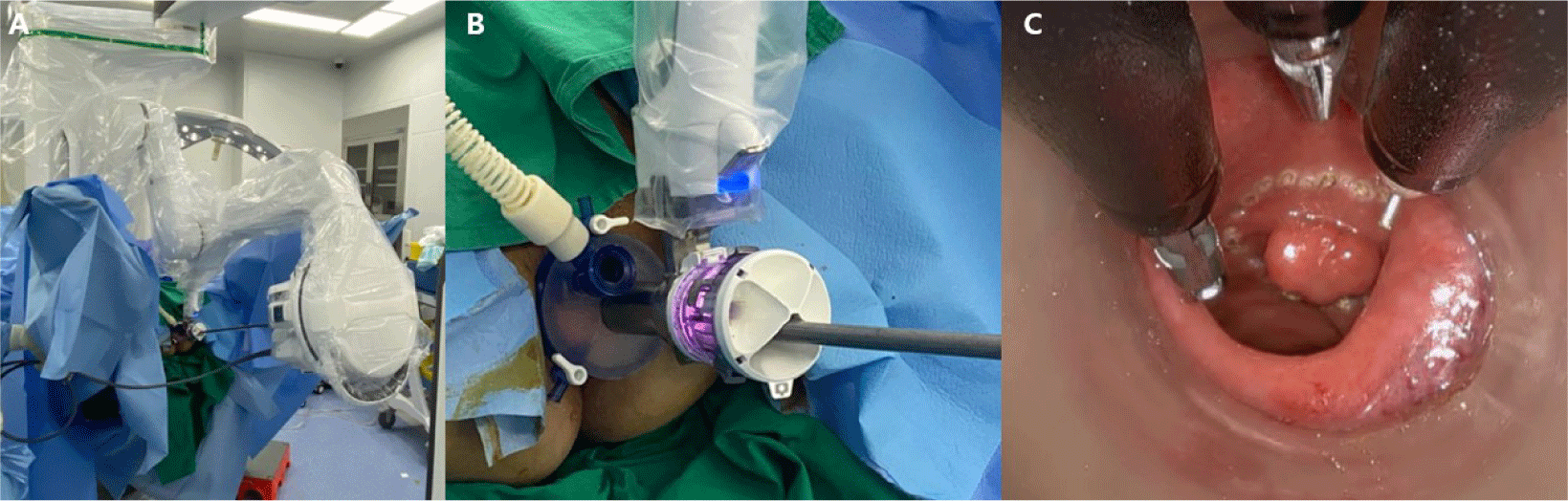
More recently, the introduction of robot-assisted TAMIS (rTAMIS), initially described in 2013, has incorporated the advantages of robotic surgery into the traditional TAMIS approach by utilizing a single access port [26]. Robotic surgery in this context offers improved ergonomics and operator dexterity through the use of articulated instruments. Additionally, it enhances image acquisition with a three-dimensional magnification view and surgeon-controlled camera, resulting in higher-quality and more stable images than can be achieved in conventional TAMIS procedures [27]. Motion scaling and tremor reduction further enhance precision, which is particularly beneficial in the constrained rectal environment. Moreover, rTAMIS demonstrates advantages in terms of ease of suturing and excision aggressiveness, especially in the upper rectal regions [28]. Another notable advantage of rTAMIS is the ability to maintain pneumo-rectum due to lower torque at the ports, as opposed to conventional TAMIS [29]. However, it should be noted that rTAMIS faces limitations related to robotic arms colliding within the narrow working area and challenges associated with lengthy docking times [30]. To address these issues, the introduction of the da Vinci SP robotic system in 2018, specifically designed for single-port use, has proven to be a significant advancement (Fig. 1) [31]. This single-port robotic platform employs a single 25 mm cannula, housing a surgeon-controlled 3D camera and three double-jointed articulated arms. Importantly, the platform offers 360° rotation of the robotic boom and instruments, enabling access to all rectal quadrants without necessitating patient or robot repositioning [32]. The availability of three arms is advantageous, as the third arm can be utilized for tissue retraction and applying suture tension during defect closure.
Outcomes of Transanal Local Excision
Postoperative complications following LE are relatively rare and are less frequent than after TME. Moreover, LE demonstrates a significant advantage over TME with respect to parameters such as length of hospital stay, incidence of postoperative complications, and the occurrence of bleeding [33]. Postoperative complications following TAE predominantly include bleeding, which is the most prevalent, along with rectal stenosis, urinary retention, fecal incontinence, and the development of rectovaginal fistulas [34]. The prevalent post-procedural complications following TEM and TAMIS procedures are bleeding, urinary tract infections, and suture line dehiscence [22].
Comparing surgical outcomes among the various operative techniques (namely, TAE, TEM, and TAMI), no statistically significant disparities were observed in terms of overall postoperative morbidity [35]. Although minimally invasive approaches tend to be associated with shorter hospital stays, statistical significance has not been achieved [35]. Moreover, no statistically significant differences were found in the duration of surgical procedures according to the surgical approach used [35]. The incidence of anorectal dysfunction following TEM and TEO procedures can be partially attributed to the utilization of a rectoscope with a 4 cm diameter, which may impact the dilatation of the anal sphincter complex. Nevertheless, it is noteworthy that the reported occurrence of fecal incontinence subsequent to the insertion of the resectoscope is 1%, and this complication is typically transient [36]. Furthermore, a systematic review assessing functional outcomes and quality of life after TEM and TAMIS observed that neither technique exhibited a significant impact on continence, with exceptions observed only in a minority of instances [37].
In the context of the learning curve, it is evident that TAMIS exhibits a comparatively shorter trajectory, potentially attributable to the pre-existing familiarity of surgeons with laparoscopic and single-access port laparoscopic techniques, unlike TEM [38]. A cohort analysis revealed that the learning curve for experienced colorectal experts in TEM was estimated to be 36 cases [36]. Additionally, the learning curve cutoff for TAMIS has been reported to be significantly shorter, ranging from 12 to 24 cases [38].
In an assessment of complication rates among patients undergoing rTAMIS, it was observed that 10.5% experienced complications. When comparing short-term outcomes between rTAMIS and conventional laparoscopic TAMIS, no statistically significant differences were detected, with the exception of an increase in procedural costs [39].
In the context of pathological outcomes, conventional TAE exhibits a notably higher positive resection margin rate (10%), which stands in stark contrast to TEM and TAMIS [25,40]. Moreover, it is essential to highlight that TAE is associated with significantly higher rates of specimen fragmentation than TAMIS procedures [35]. Specifically, the incidence of positive margins following TAMIS was reported to be 4.4%, with a concomitant tumor fragmentation rate of 4.1% [22]. This disparity may be attributed to suboptimal visualization and the utilization of non-ergonomic instruments during TAE procedures. Importantly, it is worth noting that attempts to identify differences in resection quality between TAMIS and TEM have yielded no significant differences [41].
In the context of rTAMIS, a study revealed a positive resection margin rate of 3.7%, demonstrating a modest decrease compared to the corresponding rates observed in conventional laparoscopic TAMIS, where positive resection margins typically range from 7% to 8.6% [39,42]. Lesion fragmentation was observed in 0.9% of rTAMIS cases, a rate lower than that of 5% reported for conventional TAMIS [39]. Additionally, it is worth noting that rTAMIS demonstrated a higher R0 resection rate (94.74%) than the conventional approach (90.48%), although this difference did not reach statistical significance [28].
Numerous studies have consistently reported that the incidence of postoperative local recurrence after LE for T1 rectal cancer typically ranges from 4% to 24%, whereas after TME, it is typically 0% to 7% [5,43-45]. When contrasting the outcomes of LE with those of TME, there is a considerably higher local recurrence rate among patients with T1 (ranging from 8.2% to 23%) and T2 rectal cancer (ranging from 13% to 30%) who undergo LE, as opposed to those who undergo TME for T1-T2 disease (ranging from 3% to 7.2%) [7,46,47]. A recent meta-analysis comparing TEM and TME for T1 rectal cancer found that the incidence of local recurrence following TEM was significantly greater than after TME [8]. Nevertheless, a study assessing the prognosis after LE did not find a notable difference in disease-free survival (DFS) compared to TME. Among individuals undergoing LE for T1-T2 disease, the 5-year DFS rates ranged from 55% to 93%, which was comparable to patients undergoing TME, who had a 5-year DFS rate of 77%−97% [46,48].
Comparing oncologic outcomes among various LE techniques, TEM exhibited a notably lower local recurrence rate than TAE [25]. This disparity in recurrence rates can primarily be attributed to the enhanced visibility achieved through TEM. A comparative analysis between TAE, TEM, and TAMIS found that TEM and TAMIS exhibited a lower recurrence rate when contrasted with TAE, while no significant difference in recurrence rates was identified between TEM and TAMIS [35]. In the context of rTAMIS, the observed local recurrence rate of 4.1% closely approximated the corresponding rate of 6% observed in conventional laparoscopic TAMIS [22,39].
A recent meta-analysis analyzed local recurrence rates in patients with T1 and T2 rectal cancers who underwent LE [49]. The study revealed that T1 lesions exhibited an 8.1% local recurrence rate. Subsequent subgroup analysis focused on low-risk T1 tumors, characterized by the absence of lymphovascular invasion, poor differentiation, deep submucosal invasion, tumor budding, or positive resection margins, and found a lower recurrence rate of 6.7%. Conversely, high-risk T1 lesions, defined by the presence of one or more high-risk features, displayed a higher local recurrence rate of 13.6%. In contrast, T2 tumors exhibited a notably higher local recurrence rate of 28.9%. Additionally, a predictive model estimated 5-year local recurrence rates of 18.6% for pT1 lesions and 29.3% for pT2 lesions [50]. Notably, independent predictors of local recurrence encompassed depth of invasion, increasing tumor size diameter, lymphovascular invasion, and tumor differentiation status.
Conclusion
Multiple techniques currently exist for the minimally invasive LE of early rectal cancer, each possessing distinct advantages. Despite the heightened risk of local recurrence, a less invasive procedure linked to significantly reduced morbidity and mortality, as well as enhanced functional outcomes, may hold appeal for certain patients. Therefore, it is imperative to provide thorough counseling to enable informed decision-making. The proposition of a minimally invasive procedure as an oncological compromise, yet still offering a substantial chance of cure, could be considered for a subset of patients burdened with substantial co-morbidities and limited physiological reserves, rendering them otherwise unsuitable candidates for conventional TME surgery.