

 , Boeun Lee
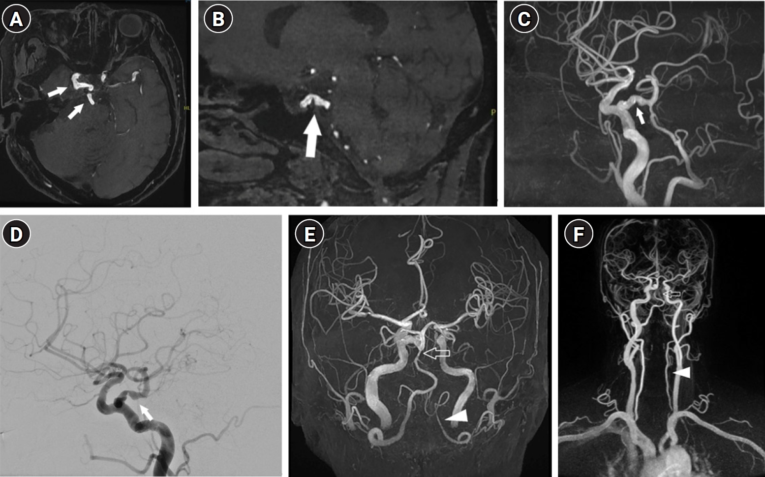
 , Muhammed Said Aydin, Gokay Ozler, Onur Ozvurmaz, Celal Kahraman, Hasan Can Çoban, Mert Kahraman, Ozan Dağdelen , Min Hyouk Beak, Won-joong Kim
 Although sciatica is commonly associated with lumbar spinal issues, it is important to acknowledge that non-spinal factors can also play a significant role in this condition. This is particularly relevant for female patients, in whom gynecologic conditions can lead to secondary sciatic neuropathy. Herein, we report the case of a 66-year-old woman who experienced posterolateral right lower extremity radiating pain. We initially performed a lumbar transforaminal epidural steroid injection, but the pain persisted. Subsequently, hip MRI revealed sciatic neuropathy adjacent to the pedunculated portions of a uterine myoma. We then performed a sub-gluteal sciatic nerve block under ultrasound guidance, resulting in significant relief of her pain. In conclusion, hip MRI can be helpful for the differential diagnosis of sciatica, and ultrasound-guided sciatic nerve block can be considered an appropriate and effective treatment option.
Citations
, Yoon Jin Choi, Ji Yeon Byun, You Won Choi, Joo Young Roh, Hae Young Choi Nontuberculous mycobacterial infections, which are often acquired from environmental sources such as water and soil, exhibit a variety of cutaneous manifestations that frequently lead to misdiagnoses and delays in treatment. A 77-year-old woman presented with multiple skin lesions in a sporotricoid distribution on her right leg, which persisted despite standard antibiotic treatments. Based on the skin biopsy, revealing granulomatous inflammation with acid-fast bacilli, and PCR testing, a nontuberculous mycobacterial infection was diagnosed. Antimycobacterial drug combinations, including clarithromycin, isoniazid, and rifampicin for 4 months, complete the skin lesion's clearance. This case underscores the need for heightened suspicion and the use of appropriate diagnostic techniques, including tissue biopsies and molecular methods such as PCR.
Citations
, Dong Chan Joo, Moon Won Lee, Bong Eun Lee, Kyungbin Kim Esophageal subepithelial tumors (SETs) are commonly encountered during screening endoscopy, and leiomyomas are the most common SET of the esophagus. Almost all patients with esophageal leiomyomas are asymptomatic; however, some present with dysphagia, depending on the size of the tumor and the extent to which it encroaches on the lumen. The typical endosonographic features of esophageal leiomyomas include well-demarcated, homogeneously hypoechoic lesions with echogenicity similar to that of the surrounding proper muscle layer, but without cystic changes. Histopathologically, esophageal leiomyomas do not undergo cystic or myxoid degeneration. This report presents a case involving a 65-year-old man with a symptomatic esophageal SET and endosonographic features indicative of malignant neoplasms, who was diagnosed with esophageal leiomyoma with cystic and myxoid degeneration following surgical resection.
Citations
Severe acute respiratory syndrome coronavirus 2 (SARS-CoV-2), the cause of coronavirus disease 2019 (COVID-19), is a type of human coronavirus that causes severe pneumonia, similar to SARS-CoV-1 and Middle East respiratory syndrome coronavirus. In Korea, the SARS-CoV-2 testing has started quickly from February 2020 to respond to the COVID-19 pandemic. In this article, I would like to introduce the characteristics of coronavirus and PCR test methods that play a large role in COVID-19 quarantine measures. Real-time reverse transcription (RT)-PCR is one of the molecular diagnostic method, and it detect SARS-CoV-2 RNA by amplifying SARS-CoV-2 specific
Citations
, Yi Kyung Kim, Ji Eun Ban, Sejung Sohn, Young Mi Hong Adenovirus infection, which has been known to mimic Kawasaki disease (KD), is one of the most frequent conditions observed during differential diagnosis when considering KD. Accordingly, it is essential to being able to differentiate between these two diseases. Therefore, we performed multiplex reverse transcriptase- polymerase chain reaction and tissue-Doppler echocardiography to distinguish between adenovirus patients and KD patients.
A total of 113 adenoviral infection patients (female 48, male 65) diagnosed from January 2010 to June 2016 were evaluated. We divided adenoviral infection patients into two groups: group 1, which consisted of individuals diagnosed with KD according to the KD American Heart Association criteria (n=62, KD with adenovirus infection); and group 2, which comprised individuals only diagnosed with adenovirus infection (n=51). Laboratory data were obtained from each patient including N-terminal pro-brain natriuretic peptide. Echocardiographic measurements were compared between two groups. In addition, reverse transcriptase-polymerase chain reaction was performed using nasopharyngeal secretions to diagnose adenoviral infection.
Conjunctival injection, cervical lymphadenopathy, polymorphous skin rash, abnormalities of the lip or oral mucosa and abnormalities of extremities were significantly higher in group 1 than group 2. Moreover, group 1 had significantly higher C-reactive protein and alanine aminotransferase levels, as well as lower platelet counts and albumin levels than group 2. Coronary artery diameter was significantly greater in group 1 than group 2.
In patients with adenoviral infection with unexplained prolonged fever, echocardiography and C-reactive protein can be used to differentiate KD with adenoviral infection from adenoviral infection alone.
, Hee Won Kang, Young Min Youn, So-Yeon Shim, Eun Ae Park, Su Jin Cho To compare the epidemiology, clinical presentation, laboratory findings, seasonality and hospital course of enteroviral meningitis (EM) and non-enteroviral meningitis (NEM) cases in infants under 3 months of age.
A retrospective chart review was performed of infants under 3 months of age or less with viral meningitis admitted to Ewha Womans University Mokdong Hospital between January 2010 and December 2016.
EM patients were more likely to have siblings compared with NEM. Most of EM was diagnosed during the summer season. Almost 80% of EM was diagnosed between July and September. Fever lasted longer in EM patients compared to NEM. White blood cell count (WBC) from the cerebrospinal fluid was higher in EM patients compared with NEM patients. WBC in blood were lower in EM patients compared with NEM patients. C-reactive protein was lower in EM patients compared with NEM patients. Most of the patients were initially started on antibiotics therapy to rule out bacterial meningitis. EM patients received shorter duration of antibiotic treatment compared with NEM patients.
This study was conducted to augment the understanding of the incidence, epidemiology, transmission in infants, clinical presentation, laboratory findings, seasonality and hospital courses of enteroviral meningitis compared to NEM. Early recognition, rapid diagnosis and proper clinical management can reduce duration of antibiotic treatment.
Citations
, Sae Han Kang, Byung Wook Jung, Hyeon Sik Oh, Min Ja Kim, Seung Hyeun Lee Differential diagnosis of invasive aspergillosis from other pulmonary fungal infections including mucormycosis is important because the treatment is pathogen-dependent. Clinically, invasive aspergillosis is often discriminated from other mold infections on the basis of typical histopathologic features in the biopsy specimen. However, biopsy alone is not always complete because different fungal species can display similar histopathologic features. Surrogate markers or molecular-based assays can be useful when the results of conventional diagnostic modalities are conflicting. Here, we present a case of invasive pulmonary aspergillosis histologically mimicking mucormycosis, which was confirmed by fungal polymerase chain reaction.
Citations
, Jeong Hyun Yoo, Jeong Soo Suh, Chung Sik Rhee Ultrasound has been found to be accurate, reliable and noninvasive method in the measurement of spleen. The study was undertaken to obtain standard values of size in three dimensions and normal range of splenic volume by the use of splenic volumetric index(SVI) in normal korean adults.
We experienced 100 cases of abdominal ultrasonography of normal korean adults from May 1995 to August 1995.
1) The average size of spleen in adult male was 6.85±1.31cm in breadth, 4.93±1.27cm in thickness, 6.33±1.46cm in height ; in adult females, 6.61±1.23cm, 5.17±1.26cm, 6.33. 42cm, respectively ; total average, 6.73±1,27cm, 5.05±1.27cm, 6.33±1.39cm,respectively.
2) The average splenic volumetric index in adult male was 8.20±3.95; in adult females, 8.41±4.08 ; total average,8.31±4.00. There were no statistical differences of SVI and size between sex and age.
Although ultrasonography is less accurate than computed tomography, it is rapid and simple method for splenic measurement.
Percutaneous biopsy is the most frequent interventional radiologic procedure. Itsincreased use is related to new imaging technique facilitating needle placement, greater safety offine needle and advances in cytology. Over a period of recent 3 years, 174 cases who underwent percutaneous needle aspiration and biopsy were analyzed.
174 biopsies under fluoroscopic or ultrasonic guidance were performed. Various anatormic sites were targeted, chest 82, liver 55, neck 10, pancreas 10, intraabdominal 6, retrperitoneal 4, thyroid 2, kidney 1, breast 1.
Obtained cytologic specimen and tissue were diagnostic in 170 of the 174 biopsies(97.7%). 4 biopsies yielded inadequate or were composed of necrotic debris. The overall accuracy for both suspected malignant and infectious diseases were 98%. The diagnosis weremalignancy in 127 biopsies and benign disease in 47 biopsies. No complications other thanpneumothorax(7 cases ; 4.1%) and a transient hemoptysis(3 cases: 17%) was observed in 10cases(5.8%).
The author obtained extremely high diagnostic accuracy of malignant andbeign or inflammatory lesions using percutaneous fine needle aspiration biopsy without seriouscomplirations. It is a valuabel diagnostic methd in the lesion of the body at any location.
, Si-Hoon Park, Gil Ja Shin, Woo Hyung Lee Femoral pseudoaneurysm is important complication after diagnostic femoral catheterizationor more complex procedure.
With the increasing use of larger-size percutaneous instruments and periprocedual anticoagulant or antiplatelet agent the incidence of postcatheterization femorl artery injuries ncluding pseudoaneuiysm has increased in the past few years.
Duplex ultrasonography and addition of color- flow Doppler provides an accurate, noninvasive. risk-free diagnosis and faster detection of intraaneurysrnal blood flow and the track betweenthe injured artery and the pseudoaneurysm.
Though early surgical repair of the arterial defect is usually recommended because of severeand life-threatening complication such as rupture, fhrornboembolism, compression neuropathyetc, Ultrasono-Guided Compression Repair(UGCR) is to be first-line treatment for its advantagesuch as high success rate, low morbidity and cost-effectiveness.
The authors report 2 cases of femoral psoudoaneurysrns treated using UGCR with nlanualcompression with C-clamp at the same time as a nonsurgical treatment.
, Eun Cheol Chung, Jeong Soo Suh, Chung Sik Rhee Abdominal ultrasound for the health screen was performed in 4610 adults from the Jan. 1993 to Mar. 1995 at Ewha University Hospital Health Clinic. Gross abnormalities were noted in the 33.3% of examined persons. The most common finding was fatty liver(21.6%). And other abnormalities were renal cyst, gallbladder stone, hepatic cyst, and hepatic calcification in the order of frequency. It is concluded that abdominal ultrasound is an important screening modality in the adults.
, Kyung Seon Park, Yoo A Choi, Ji Hee Kim, Bu Seok Jeon, Sung Ho Her Arterial remodeling is commonly observed in human atherosclerosis. It is a heterogeneous response ranging from positive remodeling to negative remodeling. Negative remodeling is a condition in which the vessel area decreases in size, often as a result of a structural change in the coronary vessel wall. But its contribution to myocardial ischemia in a de novo lesion has not been clearly shown. A 51-year-old female with exertional angina was admitted to our hospital. Coronary angiography was performed, revealing a severe stenosis at the middle part of the right coronary artery (RCA). Although we predilated ballooning at the middle RCA, the degree of stenosis did not improve. Thus intravascular ultrasound (IVUS) was performed. The lesion was not nearly showed plaque burden and severe negative remodeling. Though the cross-sectional narrowing percentage was significant, we decided to medical treatment for fearing coronary perforation by stenting. This case report intends to emphasize that severe coronary stenosis should be performed IVUS before the stenting. We describe a rare case with severe negative remodeling at the middle part of the RCA without atheroma plaque.
Citations
, Hye-Young Choi, Chung Sik Rhee, Sun Hee Sung To evaluate pathologic findings of fibrocystic disease correlated with sonographic findings in the patients with solid lesion on ultrasonography.
Total 63 pathologically proven fibrocystic disease in 57 patients are retrospectively evaluated. On ultrasonography, the lesions were divided into solid and non-solid mass-like lesions. We analyzed the margin and echogenicity of solid mass-like lesions that were correlated with pathologic findings and also statistically analyzed Chi-square and Fisher's exact test.
Ultrasonogram of fibrocystic disease showed solid mass-like lesion in 73% and non solid mass-like lesion in 27%. Among the solid lesions, well-defined margin revealed in 72%, ill-defined margin in 28% and hypoechoic in 59%, isoechoic 41%. On the pathologic analysis, the solid and the non-solid mass-like lesion showed respectively : fibrous stroma in 56.5% and 53%, fibroadenomatous change in 50% and 12%, mixed stroma in 41% and 35.3%, cystic change in 37% and 70.6%, ductectasia in 28% and 58.8%, lobular hyperplasia in 26% and 12%, ductal hyperplasia 13% and 5.9%, and adenosis in 8.7% and 0%. The solid lesions showed more fibroadenomatous change and the difference between there was statistically significant(p=0.008).
The solid mass-like lesion, which represented as a well-defined isoechoic benign mass on ultrasonogram was more common than as expected, and this was due to the fibroadenomaous change on histopathology.
, Jeong Soo Suh, Chung Sik Rhee To investigate whether measurements of hepatic metastases before contrast administration are different from measuments after contrast administration. And to gain more effective follow up method by analyzing the difference of contrast between pre- and postcontrast scans.
Thirty patients with herpatic metastases were underwent conventional CT. Continuous 10mm thick slices were obtained from liver dome to pelvic inlet, then the patients received IV injection of contrast material, and same method as precontrast CT scan was performed. Additional 5mm thin slice scan was obtained in case of need. Three radiologists performed independent bidimensional measurements of the randomly selected lesion on both pre- and postcontrast images at the same level and analyzed the difference of the size and contrast.
The size of hepatic metastases were measured as smaller on postcontrast images ; average 41.4±43.5cm2 on precontrast scan & 35.2±37.5cm2 on postcontrast scan. There was significant difference by paired t-test(p<0.02). 24 of 30 cases(80%) showed better conspicuity on postcontrast images, 5(16.7%), on precontrast images and 1(3.3%) showed similiar conspicuity on both pre- and postcontrast images. The contrast of hepatic metastases was significantly higher on postcontrast scan by chi-square test(p<0.01).
Hepatic metastases are significantly smaller on postcontrast images. The contrast between metastatic lesion & liver parenchyme was better on postcontrast scan. Therefore, serial assessment of hepatic metastases size by CT should not be compared mixed pre- and postcontrast image. And postcontrast scan is more effective method than precontrast for follow up of hepatic metastasis.
, Ku Yong Chung, Jeong Soo Suh, Chung Sik Rhee Our purpose was to discuss the current results of renal transplantation at our institute and to document the usefulness of the ultrasonography in the follow-up of renal allograft.
Thirty five renal allografts who operated and followed-up at our hospital were included. All patients underwent renal duplex and Doppler sonography. According the clinical course of allograft, the sonographic findings were classified into successful renal transplantation(SRT), acute rejection(AR), chronic rejection(CR), and graft failure(GF). We retrogradely analyzed the sonographic findings as follows : renal size(length, width, thickness), cortex echogenicity, corticomedullary differentiation, renal sinus and pyramid, renal pelvis, resistive index(RI).
Results of allografts were as follows : SRT, 24 case(68.6%) ; AR, 6(17.1%) : CR, 3(8.6%) ; and GF, 2(5.7%). The changes of length of allografts were shown no statistically significant changes between the groups, but there is significant increase of thickness of allograft in AC and GF with significance. The mean RI was statistically increased in AR(RI=0.87), and the mean RI's of other groups were 0.65, 0.70, and 0.67 in order to SRT, CR, GF. Parenchymal echogenicities are changed in 66.7% of AC and CR, 25% of SRT, and 50% of GF without clinical significance. There are changes of CMJ, pyramid, sinus echo, renal pelvis of allografts, however, which were shown no statistical significance.
Even though we have small cases and short experiences of renal transplantation at our institute, we considered we have relatively good results and it was guessed there were many efforts for the renal transplantation. The duplex and Doppler sonography were useful tools in the follow-up of allograft, especially deciding acute rejection and graft failure, although it is difficult to decide chronic rejection and can not used to differentiate between the main parenchymal causes of graft failure.
, Hee Ok Kim, Mi Young Park, Young Ju Kim, Jung Ja Ahn, Bock Hi Woo Neural tube defects are a heterogenous group of malformations resulting from failure of neural tube closure during early embryogenesis. Anencephaly is the commonest form of neural tube defect and results from failure of closure of the anterior portion of the neural tube. Anencephaly is characterized by absence of the cranium along with cerebral hemispheres that are rudimentary or absent and risk of recurrence after affected child is 2-3%. Periconceptual folic acid intake may decrease the incidence and recurrence of anencephaly.
Most often, anencephaly is discovered by conventional two-dimensional ultrasonography at the time of attempted biparietal diameter determination for fetal age in the second trimester. Two-dimensional transvaginal ultrasonography has a limitation in a motion of the transducer shaft due to narrow space of the vagina. It is sometimes impossible to obtain information of the whole brain and to miss the fetal CNS(central nervous system) anomalies. Recent advanced three-dimensional ultrasonography has remarkably improved not only surface rendering but also multiplanar analysis of internal structure.
Recently, we encountered one case of recurrent anencephaly that had occurred in a same pregnant woman and three-dimensional transvaginal ultrasonography enabled us to diagnose anencephaly at 113 weeks of gestation. We report this case with brief review of the literatures.
This study was performed to evaluate the impact of various peri-transplant factors on transfusion requirements in 45 patients with leukemia or severe aplastic anemia undergoing HLA-matched allogeneic bone marrow transplantation(BMT).
All patients were treated in an isolated room with HEPA filtration, and the combination of cyclosporin and short-course of methotrexate was used for GVHD prophylaxis. Patients received irradiated packed red cells to maintain the hematocrit ≥30% and irradiated platelet pheresis to keep the platelet count ≥20,000/µl.
In the first month, the mean(range) number of red cells and platelet pheresis were 4.9(0-21), 26.7(8-61), respectively. On univariate analyses, pre-BMT status(high-risk : 7.94±5.14 vs standard-risk: 3.78±2.99, p=0.0076) and concurrent infection(present : 8.41±4.70 vs absent : 3.33±2.72, p=0.0005) and sex incompatibility(match : 4.67±3.72 vs female → male : 3.78±3.07 vs male → female : 9.13±5.74, p=0.0161) were significantly associated with red cell requirements in the first month. Also, high-risk pre-BMT status(32.25±16.15 vs 20.25±14.64, p=0.0l56), the presence of concurrent infection(39.35±16.42 vs 15.33±5.67, p=0.0001) and veno-occlusive disease(45.00±14.47 vs 22.00±14.49, p=0.0055) increased platelet requirements significantly after allogeneic BMT. In particular, pre-BMT disease status was found to be independently associated with transfusion requirements.
This study demonstrates that pre-BMT status does influence transfusion requirements in the first month after HLA-matched allogeneic BMT. Further studies are necessary to confirm these results and to define optimal transfusion strategies.
, Kyung Hee Kim To access the changes of cerebral blood flow velocity according to the time after surfactant administration, we prospectively studied in the Hyaline Membrane Disease using Doppler ultrasonography.
The patients were 26 infants. The mean gestational age was 3l4wks (range, 184 to 38wk). The ratio of male : female was 16 : 10, mean weight was 1.76±0.88Kg, Apgar score at 5min was 6.9, and type of delivery was C-section : vaginal delivery 19 : 4. Before and after, 10, 30min, 1, 6, l2hr, 1, 3, 5, 7days after surfactant administration, peak systolic and end-diastolic flow velocity(PSFV, EDFV) were estimated by Doppler method measuring MCA flow velocity. The Resistive index was calculated according to the mathematics. For the evaluation of the clinical status, systolic and diastolic systemic BP, PaO2, PaCO2, FiO2, pH, and respiratory rate(RR) were checked.
The cerebral blood flow velocity showed initial increase of PSFV just after synthetic surfactant administration, and the increased PSFV continued until the 30 minites and then decreased. PSFV returns to initial level at 6hr, and then increased again. The changes of EDFV was not significant. The changes of RI & PI were no significant changes. The effects of surfactant to the systemic BP had no significance. The changes of PaCO2 and PaO2 were not significant. FiO2 showed steady improvement. Initial tachypnea and acidosis progressively improved without clinical significance.
The administration of Surfactant in the HMD patients results in transient increase of cerebral blood flow velocity.
, Duk Hee Kang, Gyu Bok Choi, Kyun Ill Yoon Patients undergoing maintenance hemodialysis(HD) potentially have an increased risk of exposure to viral hepatitis. The reported prevalence of antiHCV in hemodialysis patients varied widely form 7.6-54% according to dialysis center and there were there were many reports that showed the correlation between the prevalence of antiHCV and duration of HD or transfusion amount.
Fifty-four patients on regular hemodialysis at our hospital were evaluated for the presence of hepatitic C antibody(antiHCV) with the comparison of various parameters such as duration of HD, amount of transfusion, past history of hepatitis, serologic markers of hepatitis B and current liver function. AntiHCV using second-generation enzyme linked immunosorbant assay were found in six of 54HD patients(11.1%). Among six antiHCV(+) percent four patients were found to have HCV-RNA in their plasma detected by PCR. The percent of male patients were significantly higher in antiHCV(+) group(66.7 vs 31.3%, p<0.05). The positivity of antiHCV did not correlated with the duration of HD and amount of transfusion(p>0.05), but prevalence increased over 2 years (5.9% in 1991, 11.1% in 1993) and HBsAg prevalence remained unchanged(9.8% in 1991, 9.3% in 1993).
Therefore, regular follow-up of liver function test and use of separate machine for antiHCV positive patients may be needed to prevent the transmission of the hepatitis C virus during the hemodialysis process itself.
 First
First Prev
Prev

