• Contact us
  • E-Submission
ABOUT
BROWSE ARTICLES
JOURNAL POLICIES
FOR CONTRIBUTORS

Articles

Page Path

Case Report

Hypocitraturia-related Ureteral Steinstrasse in a Renal Transplant Recipient

The Ewha Medical Journal 2015;38(3):117-120. Published online: October 31, 2015

Department of Internal Medicine, Samsung Changwon Hospital, Sungkyunkwan University School of Medicine, Changwon, Korea.

Corresponding author: Yu-Ji Lee. Department of Internal Medicine, Samsung Changwon Hospital, Sungkyunkwan University School of Medicine, 158 Paryong-ro, Masanhoewon-gu, Changwon 51353, Korea. Tel: 82-55-290-6332, Fax: 82-55-290-1064, yuji.lee@samsung.com
• Received: May 16, 2015   • Accepted: August 20, 2015

Copyright © 2015, The Ewha Medical Journal

This is an Open Access article distributed under the terms of the Creative Commons Attribution Non-Commercial License (http://creativecommons.org/licenses/by-nc/3.0/) which permits unrestricted non-commercial use, distribution, and reproduction in any medium, provided the original work is properly cited.

  • 59 Views
  • 0 Download
prev next
  • Urolithiasis is an uncommon complication in renal transplantation. We report a case of hypocitraturia-related ureteral steinstrasse which was spontaneously formed in a renal transplant recipient. The patient who underwent renal transplantation was admitted with acute pyelonephritis. Hydronephrosis in the transplanted kidney and multiple stones (steinstrasse) in the distal ureter were incidentally found on computed tomography scanning. After a failed attempt of ureteroscopic removal of stones, the patient underwent open ureterolithotomy and ureteroureterostomy. On stone analysis, carbonate apatite was confirmed. Urinary citric acid levels were decreased to 127.6 mg/day. Potassium citrate was administered to prevent stone recurrence by increasing urinary citrate excretion. No recurrence of stones was shown six months later. Urolithiasis in renal transplant recipients requires a high index of suspicion. Hypocitraturia can increase the risk for urolithiasis. Rapid recognition by careful surveillance, prompt removal of stones, and precautionary efforts to prevent recurrence are needed.
  • 1. Mamarelis G, Vernadakis S, Moris D, Altanis N, Perdikouli M, Stravodimos K, et al. Lithiasis of the renal allograft, a rare urological complication following renal transplantation: a single-center experience of 2,045 renal transplantations. Transplant Proc 2014;46:3203-3205.
  • 2. Ferreira Cassini M, Cologna AJ, Ferreira Andrade M, Lima GJ, Medeiros Albuquerque U, Pereira Martins AC, et al. Lithiasis in 1,313 kidney transplants: incidence, diagnosis, and management. Transplant Proc 2012;44:2373-2375.
  • 3. Kim H, Cheigh JS, Ham HW. Urinary stones following renal transplantation. Korean J Intern Med 2001;16:118-122.
  • 4. Verrier C, Bessede T, Hajj P, Aoubid L, Eschwege P, Benoit G. Decrease in and management of urolithiasis after kidney transplantation. J Urol 2012;187:1651-1655.
  • 5. Salem S, Mehrsai A, Zartab H, Shahdadi N, Pourmand G. Complications and outcomes following extracorporeal shock wave lithotripsy: a prospective study of 3,241 patients. Urol Res 2010;38:135-142.
  • 6. Harper JM, Samuell CT, Hallson PC, Wood SM, Mansell MA. Risk factors for calculus formation in patients with renal transplants. Br J Urol 1994;74:147-150.
  • 7. Stapenhorst L, Sassen R, Beck B, Laube N, Hesse A, Hoppe B. Hypocitraturia as a risk factor for nephrocalcinosis after kidney transplantation. Pediatr Nephrol 2005;20:652-656.
  • 8. Hamm LL, Hering-Smith KS. Pathophysiology of hypocitraturic nephrolithiasis. Endocrinol Metab Clin North Am 2002;31:885-893.
  • 9. Hamm LL. Renal handling of citrate. Kidney Int 1990;38:728-735.
  • 10. Simpson DP. Citrate excretion: a window on renal metabolism. Am J Physiol 1983;244:F223-F234.
  • 11. Cicerello E, Merlo F, Mangano M, Cova G, Maccatrozzo L. Urolithiasis in renal transplantation: diagnosis and management. Arch Ital Urol Androl 2014;86:257-260.
  • 12. Homayoon K. Spontaneous steinstrasse due to renal tubular acidosis. Br J Urol 1996;77:610-611.
  • 13. Van Savage JG, Fried FA. Bilateral spontaneous steinstrasse and nephrocalcinosi associated with distal renal tubular acidosis. J Urol 1993;150(2 Pt 1):467-468.
  • 14. Stravodimos KG, Adamis S, Tyritzis S, Georgios Z, Constantinides CA. Renal transplant lithiasis: analysis of our series and review of the literature. J Endourol 2012;26:38-44.
Fig. 1

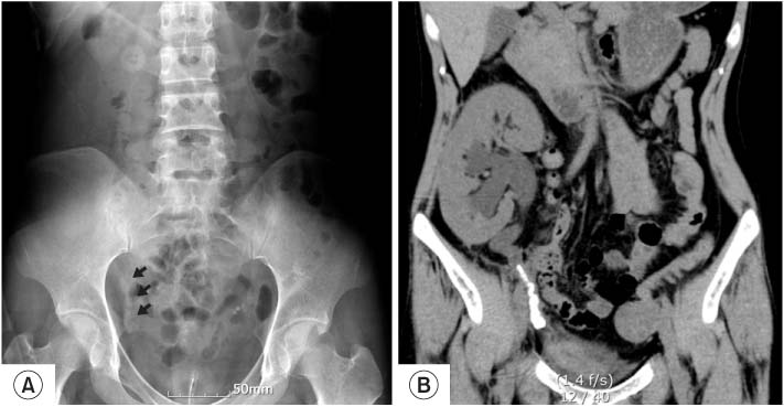
Initial radiologic findings. (A) The plain film of the abdomen reveals radiopaque stones in the pelvic cavity (arrow). (B) Computed tomography scan shows hydroureteronephrosis in transplanted kidney and multiple stones in the distal ureter (steinstrasse).

emj-38-117-g001.jpg
Fig. 2

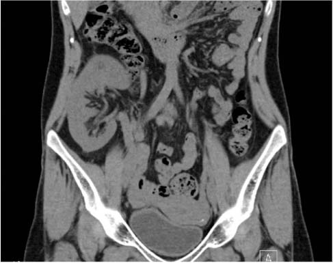
Computed tomography after six months. It show no stones in transplanted kidney and ureter.

emj-38-117-g002.jpg
Table 1

Result of Stone analysis

emj-38-117-i001.jpg

Figure & Data

References

    Citations

    Citations to this article as recorded by  

      Download Citation

      Download a citation file in RIS format that can be imported by all major citation management software, including EndNote, ProCite, RefWorks, and Reference Manager.

      Format:

      Include:

      Hypocitraturia-related Ureteral Steinstrasse in a Renal Transplant Recipient
      Ewha Med J. 2015;38(3):117-120.   Published online October 31, 2015
      Download Citation
      Download a citation file in RIS format that can be imported by all major citation management software, including EndNote, ProCite, RefWorks, and Reference Manager.

      Format:
      • RIS — For EndNote, ProCite, RefWorks, and most other reference management software
      • BibTeX — For JabRef, BibDesk, and other BibTeX-specific software
      Include:
      • Citation for the content below
      Hypocitraturia-related Ureteral Steinstrasse in a Renal Transplant Recipient
      Ewha Med J. 2015;38(3):117-120.   Published online October 31, 2015
      Close

      Figure

      • 0
      • 1
      Hypocitraturia-related Ureteral Steinstrasse in a Renal Transplant Recipient
      Image Image
      Fig. 1 Initial radiologic findings. (A) The plain film of the abdomen reveals radiopaque stones in the pelvic cavity (arrow). (B) Computed tomography scan shows hydroureteronephrosis in transplanted kidney and multiple stones in the distal ureter (steinstrasse).
      Fig. 2 Computed tomography after six months. It show no stones in transplanted kidney and ureter.
      Hypocitraturia-related Ureteral Steinstrasse in a Renal Transplant Recipient

      Result of Stone analysis

      Table 1 Result of Stone analysis

      TOP