Abstract
Primary leiomyosarcoma of the breast is an extremely rare malignancy, accounting for less than 1% of all breast tumors. Diagnosis is challenging because its morphology overlaps with that of other spindle cell lesions, and standardized treatment guidelines are currently unavailable. A 44-year-old woman presented with a rapidly enlarging, firm, 15×15 cm mass in the left breast. She had previously undergone surgery elsewhere, with diagnoses of leiomyoma and desmoid-type fibromatosis. Imaging demonstrated a lobulated mass without evidence of metastasis. Excision revealed pleomorphic spindle cells arranged in intersecting fascicles, with necrosis and dermal invasion. Primary breast leiomyosarcoma was confirmed by immunohistochemistry, which demonstrated smooth muscle actin and desmin positivity and negative staining for pancytokeratin, p63, S100, CD34, and BCL2. The patient underwent modified radical mastectomy followed by adjuvant radiotherapy and chemotherapy. Primary breast leiomyosarcoma is a rare entity that remains diagnostically challenging. Immunohistochemistry is essential for accurate diagnosis, and optimal management requires a dedicated multidisciplinary approach.
-
Keywords: Leiomyosarcoma; Spindle cell tumor; Immunohistochemistry; Case reports
Introduction
Background
Primary leiomyosarcoma of the breast is an extremely rare malignant mesenchymal tumor, comprising less than 1% of breast neoplasms. It presents substantial diagnostic and therapeutic challenges because available data are limited and standardized treatment guidelines are lacking [
1]. The tumor is thought to arise from smooth muscle cells of the nipple–areolar complex, vessel walls, or mesenchymal stem cells with smooth muscle differentiation [
2]. Patients typically present with a rapidly enlarging, painless breast mass, and the diagnosis is often established only after excisional histopathological evaluation because core biopsy and cytology may be misleading [
3].
Immunohistochemistry is essential for distinguishing leiomyosarcoma from other spindle cell lesions of the breast [
4]. Because of its rarity, treatment strategies are usually extrapolated from soft tissue sarcoma protocols [
5]. Although the prognosis may be relatively favorable, biological behavior is unpredictable, with ongoing risks of recurrence and metastasis [
6]. Reporting such cases is important for improving diagnostic accuracy and informing management of this rare tumor.
This case of primary leiomyosarcoma of the breast highlights the diagnostic challenges associated with this entity and the critical role of immunohistochemistry in establishing the correct diagnosis. This report also describes the management approach used in the absence of standardized guidelines and contributes to the understanding of the clinicopathological features and outcomes of this uncommon spindle cell tumor of the breast.
Case presentation
Ethics statement
Written informed consent was obtained from the patient for publication of this case report and the accompanying images. All patient-related information has been anonymized to protect privacy and confidentiality in accordance with institutional and international research ethics guidelines. No identifying personal data have been disclosed.
Patient information
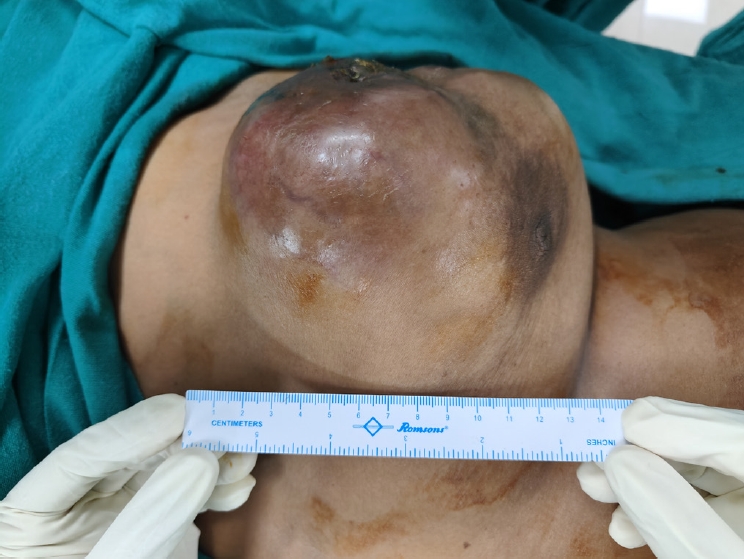
A 44-year-old woman presented with an enlarging, firm lump in the upper outer quadrant of the left breast, measuring 15×15 cm, with overlying erythema and a previous surgical scar (
Fig. 1). She had a history of recurrent breast lesions, including prior diagnoses of leiomyoma and desmoid-type fibromatosis at an outside facility; both lesions had been managed with lumpectomy. Fine-needle aspiration cytology (FNAC), also performed elsewhere, suggested proliferative breast disease with atypia and prompted further evaluation. The smears could not be reviewed.
Ultrasonography suggested a Breast Imaging Reporting and Data System category 4B lesion. Positron emission tomography–computed tomography (PET-CT), performed at an outside facility, revealed a 14.8 cm heterogeneously enhancing tumor with lobulated margins and central necrosis. The lesion closely abutted the skin but showed no invasion of the pectoral muscle or adjacent fat planes. Axillary lymph nodes showed no abnormal fluorodeoxyglucose (FDG) uptake and had preserved fatty hila, indicating no metastatic involvement. No additional FDG-avid lesions or tumor deposits were identified elsewhere in the body, suggesting no evidence of disease at other sites.
Core biopsy revealed a malignant spindle cell tumor composed of elongated cells arranged in fascicles, with moderate pleomorphism and 8–10 mitotic figures per 10 high-power fields (HPFs). The differential diagnoses included borderline or malignant phyllodes tumor, metaplastic carcinoma, and other mesenchymal tumors. Immunohistochemistry could not be performed on the biopsy specimen to further characterize the tumor.
The patient underwent left modified radical mastectomy; the resected specimen measured 24×20×11 cm. The tumor measured 15×15×10 cm, occupied a substantial portion of the breast, and was associated with overlying skin ulceration (
Fig. 2). Microscopic evaluation confirmed a malignant spindle cell tumor with infiltrative margins. The spindle cells were arranged in fascicles and showed pleomorphic, hyperchromatic, elongated nuclei, high mitotic activity (11 per 10 HPFs; 6/mm
2), and necrosis (
Fig. 3A–
D). No epithelial component was identified. The tumor invaded the dermis but showed no vascular or lymphatic invasion, and all 9 dissected lymph nodes were uninvolved.
Immunohistochemistry demonstrated positivity for smooth muscle actin (SMA) and desmin, supporting smooth muscle differentiation, whereas staining for pancytokeratin, p63, BCL2, S100, and CD34 was negative (
Fig. 4A,
B). The Ki-67 labeling index was 15% (
Fig. 4C). Based on the histopathological and immunohistochemical findings, the tumor was diagnosed as primary leiomyosarcoma of the breast, grade 2, according to the French Federation of Cancer Centers Sarcoma Group grading system.
Given the high-grade features, large tumor size (>10 cm; pT3), and moderate proliferative activity, a multimodal treatment approach was planned. Because no standardized treatment guidelines exist specifically for primary breast leiomyosarcoma, management was extrapolated from established soft tissue sarcoma protocols after multidisciplinary team discussion, consistent with current oncological practice for rare sarcomas [
1,
5]. Adjuvant radiotherapy (60 Gy in 30 fractions) was administered to the chest wall because of the close surgical margins (0.3 cm) and to reduce the risk of local recurrence. Ultrasonography performed after completion of radiotherapy showed a postoperative hypoechoic collection suggestive of a seroma, measuring 3.3×0.6 cm, at the surgical scar site, with no evidence of calcification or vascularity. Subsequently, whole-body FDG PET-CT showed FDG-avid thickening and subcutaneous fat stranding in the left chest wall, with a maximum standardized uptake value of 5.3. Close follow-up of the lesion was advised. The patient then underwent 6 cycles of doxorubicin, ifosfamide, and mesna (AIM) chemotherapy. She tolerated radiotherapy and chemotherapy well. Supportive care included mesna for uroprotection during ifosfamide therapy and granulocyte colony-stimulating factor support with pegfilgrastim or filgrastim to prevent neutropenia. Ultrasonography performed after completion of 2 chemotherapy cycles showed mild subcutaneous edema at the skin incision site and no postoperative collection. At follow-up after completion of chemotherapy, a local clinical examination showed no evidence of disease. The patient was advised to undergo PET-CT as part of follow-up assessment of disease status.
Discussion
Primary sarcomas are rare tumors that may arise in any organ; however, occurrence in the breast is exceptionally uncommon, accounting for nearly 1% of breast malignancies and fewer than 5% of all soft tissue sarcomas [
1]. The clinical presentation is often nonspecific, and most patients present with an enlarging, painless breast mass, as in the present case. Reported tumor sizes range from 0.3 to 16 cm, with a mean size of 4.7 cm [
2]. Although these tumors predominantly affect women, rare cases have also been reported in men [
3].
The tumor is believed to arise from smooth muscle cells of the lactiferous ducts, blood vessels, or erector pili muscles in the periareolar region [
4]. Secondary cases may develop after prior radiotherapy or in the setting of chronic lymphedema [
5]. Imaging modalities such as mammography and ultrasonography generally lack specificity and may lead to misclassification as fibroadenoma or phyllodes tumor, further complicating diagnosis [
6].
The patient’s previous outside diagnoses of leiomyoma and desmoid-type fibromatosis warrant consideration. Because the original slides from these lesions were not available for review, definitive retrospective reclassification was not possible. Synchronous or metachronous tumors are very rare. A literature search identified no reports of synchronous or metachronous breast spindle cell neoplasms with differing histologic diagnoses. Therefore, the prior diagnoses may have reflected sampling error or may have been rendered without immunohistochemical support.
The initial cytological evaluation in this case reportedly showed spindle-shaped cells arranged in fascicles with blunt-ended nuclei. However, FNAC has substantial diagnostic limitations in such cases because of cytomorphological overlap with other spindle cell lesions of the breast [
7]. Under the Yokohama System for Reporting Breast Fine-Needle Aspiration Biopsy Cytopathology, spindle cell tumors may be classified as “suspicious for malignancy” or “malignant,” depending on the degree of cellular atypia and the presence of mitotic activity or necrosis [
8]. The absence of epithelial elements in the smears, together with pleomorphic spindle cells in clusters, may support classification in the “malignant” category. According to the World Health Organization Reporting System for Soft Tissue Cytopathology, such a lesion may be categorized as a soft tissue neoplasm of uncertain malignant potential if only mildly pleomorphic spindle cells are observed. However, if significant nuclear atypia is present, the tumor would be categorized as “suspicious for malignancy.” If mitotic figures and/or necrosis are evident, although these features are often difficult to appreciate cytologically, the lesion would be classified as “malignant” [
9].
Several spindle cell lesions should be considered in the cytological differential diagnosis. Metaplastic carcinoma may mimic sarcoma cytologically, with pleomorphic spindle cells, vacuolated cytoplasm, and occasional epithelial clusters. Malignant phyllodes tumor, a relatively more common entity in this setting, should be carefully excluded. It typically shows atypical spindle nuclei within stromal fragments and numerous naked nuclei in a hypercellular background. In contrast, leiomyosarcoma is more likely to show cohesive clusters and fascicles of pleomorphic spindle cells. Undifferentiated pleomorphic sarcoma shows marked pleomorphism and bizarre multinucleated giant cells [
10]. Benign spindle cell lesions, such as fibromatosis, show bland, uniform spindle cells without significant pleomorphism or mitotic activity; accurate identification is therefore important to avoid overtreatment. Thus, FNAC alone is often insufficient for a definitive diagnosis, and histological confirmation by core needle or excisional biopsy, together with immunohistochemistry, is required [
11].
Core needle biopsy was subsequently performed in this case. Although core biopsy provides better architectural assessment than FNAC, diagnostic limitations remain, particularly because primary sarcomas such as leiomyosarcoma lack ductal epithelial elements. This absence can create diagnostic uncertainty and make it difficult to distinguish sarcoma from metaplastic carcinoma, which often contains an epithelial component. Therefore, correlation with definitive excisional histopathology and immunohistochemistry is essential [
12].
Histopathological examination of the surgical specimen in leiomyosarcoma typically shows intersecting fascicles of spindle cells with eosinophilic cytoplasm, elongated blunt-ended nuclei, moderate to severe nuclear atypia, brisk mitotic activity, and/or necrosis [
13]. In the present case, no lymph node involvement was identified, consistent with the general observation that nodal metastasis is uncommon in breast leiomyosarcoma [
2].
Because of overlapping histological appearances, several benign and malignant spindle cell tumors of the breast must be excluded. Fibromatosis typically shows uniform, bland spindle cells with β-catenin and SMA positivity but is negative for desmin and S100 [
14]. Nodular fasciitis is characterized by plump spindle cells in a myxoid stroma, with SMA positivity and negative β-catenin and S100 expression. Myofibroblastoma is usually well circumscribed and is immunoreactive for desmin, CD34, estrogen receptor, and progesterone receptor [
15].
Among malignant differential diagnoses, metaplastic carcinoma is particularly challenging because it may show a monophasic spindle cell pattern that histologically mimics sarcoma. It is usually positive for cytokeratin and p63 and negative for S100. Malignant phyllodes tumor demonstrates stromal overgrowth and scant epithelial elements, with CD34 and SMA positivity. Malignant peripheral nerve sheath tumor (MPNST) shows wavy spindle cells with frequent mitoses and is positive for S100 and SOX10 [
10]. In addition, neurotrophic tyrosine receptor kinase fusion-positive sarcomas may show minimal pleomorphism and fascicular architecture and require molecular confirmation because of potential therapeutic implications [
16]. In the present case, adequate sampling and immunohistochemistry were decisive in confirming the diagnosis. The tumor showed diffuse immunoreactivity for SMA and desmin, supporting smooth muscle lineage. Negative immunostaining for pancytokeratin, p63, BCL2, S100, and CD34 helped exclude metaplastic carcinoma, phyllodes tumor, and MPNST. Fibromatosis was not favored in the present case because of marked nuclear pleomorphism, necrosis, and mitotic activity.
Table 1 summarizes the key immunohistochemical profiles of the main spindle cell lesions in the differential diagnosis.
Surgical excision with clear margins remains the mainstay of treatment. Modified radical mastectomy was performed in the present case and is the most frequently adopted approach. Wide local excision may be feasible for smaller tumors. Achieving negative surgical margins is critical because it substantially reduces local recurrence rates [
1]. The roles of chemotherapy and radiotherapy are not well established because of limited evidence. However, anthracycline-based chemotherapy is often used in high-risk or metastatic cases, whereas radiotherapy may be considered for positive margins or larger tumors [
17]. In the present case, radiotherapy was selected because of the close surgical margins and large tumor size, and AIM chemotherapy was selected because of the high-grade histology and large tumor burden, following general soft tissue sarcoma management principles in the absence of breast leiomyosarcoma-specific guidelines [
1].
The prognosis of breast leiomyosarcoma is generally favorable compared with that of other soft tissue sarcomas. However, hematogenous metastasis occurs in approximately 25% of cases, most commonly involving the lungs, liver, and bones. Metastases may develop years after initial treatment, emphasizing the need for long-term surveillance [
2]. Because of the rarity of this tumor, current treatment protocols are largely extrapolated from soft tissue sarcoma guidelines, underscoring the need for additional studies and accumulated case experience to guide standardized management [
1].
In conclusion, primary leiomyosarcoma of the breast is a rare malignancy that poses significant diagnostic and therapeutic challenges. Accurate diagnosis relies on histopathological and immunohistochemical evaluation because conventional imaging and cytology may be inconclusive. Given the absence of standardized treatment guidelines, management is generally adapted from soft tissue sarcoma protocols, with complete surgical excision remaining the mainstay of treatment. Owing to the risk of recurrence and metastasis, multidisciplinary management and long-term follow-up are essential to optimize patient outcomes.
-
Authors’ contribution
AP and AR contributed to manuscript preparation and histopathological diagnosis. IP assisted in histopathological diagnosis. KR was involved in surgical management and provided clinical images. CR contributed radiation oncology expertise. All authors participated in manuscript drafting and revision, and all have read and approved the final manuscript.
-
Conflict of interest
No potential conflict of interest relevant to this article was reported.
-
Funding
No external funding was received for this study.
-
Data availability
Not applicable.
-
Acknowledgments
None.
-
Supplementary materials
None.
Fig. 1.Clinical image showing a 15×15 cm left breast mass in the upper outer quadrant with overlying erythema and a pre-existing surgical scar.

Fig. 2.Gross cut surface showing a firm, gray-white tumor with a whorled appearance.

Fig. 3.Histopathological features of primary breast leiomyosarcoma. (A) Interlacing fascicles of spindle cells (hematoxylin and eosin [H&E], ×40). (B) Pleomorphic spindle cells with hyperchromatic nuclei and nuclear atypia (H&E, ×400). (C) Atypical mitotic figure (arrow) (H&E, ×400). (D) Area of tumor necrosis with adjacent viable pleomorphic spindle cells (H&E, ×400).

Fig. 4.Immunohistochemical staining of the tumor. (A) Desmin-positive tumor cells (×400). (B) Smooth muscle actin-positive tumor cells (×400). (C) Ki-67 labeling index of 15% in tumor cells (×400).

Table 1.Comparative immunohistochemical profiles of spindle cell lesions in the differential diagnosis of primary breast leiomyosarcoma
|
Entity |
SMA |
Desmin |
h-caldesmon |
Pan-CK |
p63 |
S100 |
CD34 |
β-catenin |
|
LMS |
+ |
+ |
+ |
− |
− |
− |
− |
− |
|
Metaplastic carcinoma |
+ |
− |
− |
+ |
+ |
− |
− |
− |
|
Malignant phyllodes tumor |
V |
− |
− |
− |
− |
− |
+ |
V |
|
Fibromatosis |
+ |
− |
− |
− |
− |
− |
− |
+ (nuclear) |
|
MPNST |
− |
− |
− |
− |
− |
+ (focal) |
V |
− |
|
This case |
+ |
+ |
ND |
− |
− |
− |
− |
ND |
References
- 1. Alimi A, Abbassi H, Abid S, Dinari A, Khlifi A, Hidar S, Boughizane S, Khairi H. Leiomyosarcoma of the breast: case report and review of the literature. Eur J Breast Health 2025;21:277-280. https://doi.org/10.4274/ejbh.galenos.2025.2024-10-8
- 2. Masadah R, Anwar F, Nelwan BJ, Faruk M. Primary leiomyosarcoma of the breast: a case report and literature review. Int J Surg Case Rep 2023;106:108290. https://doi.org/10.1016/j.ijscr.2023.108290
- 3. Ely Cheikh T, Hamza K, Hicham B, Fatiha EM, Hajar EO, Mustapha B, Mohamed E, Mohamed E. Leiomyosarcoma of the male breast: case report. Ann Med Surg (Lond) 2021;67:102495. https://doi.org/10.1016/j.amsu.2021.102495
- 4. Sethi E, Misra S, Ahuja A. Primary leiomyosarcoma with osteosarcomatous differentiation of the breast. Autops Case Rep 2024;14:e2024476. https://doi.org/10.4322/acr.2024.476
- 5. Hayashi S, Kitada M, Matsuda Y, Ishibashi K, Takahashi N. Leiomyosarcoma arising in irradiated region after breast-conserving surgery: a case report. Surg Case Rep 2015;1:76. https://doi.org/10.1186/s40792-015-0072-y
- 6. Khan M, Siddiqui B, Rafey M, Hassan M, Faridi S. A rare case report of primary breast leiomyosarcoma presenting clinicoradiologically as phyllodes tumour. Indian J Pathol Oncol 2025;12:86-89. https://doi.org/10.18231/j.ijpo.2025.019
- 7. Jun Wei X, Hiotis K, Garcia R, Hummel Levine P. Leiomyosarcoma of the breast: a difficult diagnosis on fine-needle aspiration biopsy. Diagn Cytopathol 2003;29:172-178. https://doi.org/10.1002/dc.10359
- 8. Yu W, Gan Q, Gong Y. The Yokohama system for reporting breast cytopathology. J Clin Transl Pathol 2023;3:99-105. https://doi.org/10.14218/JCTP.2023.00006
- 9. Bui MM. World Health Organization Reporting System for Soft Tissue Cytopathology: a concise review with a practical diagnostic approach. J Clin Transl Pathol 2025;5:72-78. https://doi.org/10.14218/JCTP.2025.00016
- 10. Rakha EA, Brogi E, Castellano I, Quinn C. Spindle cell lesions of the breast: a diagnostic approach. Virchows Arch 2022;480:127-145. https://doi.org/10.1007/s00428-021-03162-x
- 11. Gupta RK, Kenwright D, Naran S, Lallu S, Fauck R. Fine needle aspiration cytodiagnosis of leiomyosarcoma of the breast: a case report. Acta Cytol 2000;44:1101-1105. https://doi.org/10.1159/000328606
- 12. Sandhya B, Babu V, Parthasarathy G, Kate V, Ananthakrishnan N, Krishnan R. Primary leiomyosarcoma of the breast: a case report and review of literature. Indian J Surg 2010;72:286-288. https://doi.org/10.1007/s12262-010-0092-6
- 13. Tharwani ZH, Habib Z, Ahmed Y, Ishrat S, Nashwan AJ. A rare case of high-grade spindle cell sarcoma of the breast: a case report. Case Rep Oncol 2024;17:217-224. https://doi.org/10.1159/000536125
- 14. Ni Y, Tse GM. Spindle cell lesions of the breast: a diagnostic algorithm. Arch Pathol Lab Med 2023;147:30-37. https://doi.org/10.5858/arpa.2022-0048-RA
- 15. Hou Y, Li Z. Spindle cell lesions of the breast. Hum Pathol Rep 2021;26:300565. https://doi.org/10.1016/j.hpr.2021.300565
- 16. Schwartz CJ, Ye J, Devine WP, Chen YY. NTRK-fusion spindle cell tumour in the breast: expanding the differential of challenging spindle cell lesions on core biopsy. Histopathology 2025;86:1018-1021. https://doi.org/10.1111/his.15421
- 17. Samenova D, Midlenko A, Khamzina Y, Kaldybayev M, Khamzina S. Primary leiomyosarcoma of the breast: a successful surgical treatment in a 45-year-old woman. Am J Case Rep 2023;24:e939437. https://doi.org/10.12659/AJCR.939437