Abstract
Appendiceal mucinous adenocarcinoma is a rare gastrointestinal malignancy that may metastasize to the ovaries and closely mimic advanced primary ovarian cancer, creating diagnostic and therapeutic challenges. A 61-year-old postmenopausal woman presented with postmenopausal bleeding, abdominal distension, abdominal pain, and weight loss. Imaging demonstrated bilateral adnexal masses with omental caking and ascites, suggestive of advanced ovarian malignancy. Cancer antigen 125 was elevated, and carcinoembryonic antigen (CEA) levels were markedly increased. She had a history of acute appendicitis 1 year earlier. Biopsy revealed mucinous adenocarcinoma with signet-ring cells. Endoscopic evaluation was unremarkable. Despite neoadjuvant chemotherapy for presumed ovarian cancer, the disease progressed, necessitating cytoreductive surgery. Histopathological examination demonstrated bilateral mucinous adenocarcinoma with peritoneal spread. Immunohistochemistry showed positivity for cytokeratin 20, caudal-type homeobox 2, and special AT-rich sequence-binding protein 2 and negativity for cytokeratin 7 and paired-box gene 8, supporting an appendiceal origin. She was started on fluorouracil, leucovorin, and oxaliplatin chemotherapy. Metastatic appendiceal carcinoma can closely resemble primary ovarian malignancy. Bilateral mucinous ovarian tumors with elevated CEA levels and poor chemotherapy response should prompt evaluation for a gastrointestinal primary tumor.
-
Keywords: Appendiceal carcinoma; Metastasis; Mucinous adenocarcinoma; Primary ovarian cancer; Signet-ring cells; Case reports
Introduction
Metastatic involvement of the ovaries most commonly arises from primary tumors of the gastrointestinal (GI) tract and can masquerade as primary ovarian neoplasms, creating a substantial diagnostic challenge [
1,
2]. Appendiceal tumors are a rare and heterogeneous group of neoplasms, accounting for approximately 0.9%–1.4% of all appendectomy specimens [
3]. They are often discovered incidentally during appendectomy or on subsequent histopathological evaluation. Although their exact etiology remains unclear, chronic inflammation, mucosal hyperplasia, and underlying genetic alterations have been implicated. The risk increases with advancing age, and a slight female predominance has been reported [
3].
Within this spectrum, appendiceal mucinous neoplasms represent a distinctive subtype, with ovarian metastases reported in nearly 50% of affected patients [
4]. When metastatic spread to the ovaries occurs, these lesions frequently mimic primary mucinous ovarian tumors, creating considerable diagnostic difficulty. This difficulty reflects their nonspecific clinical presentation, overlapping radiological features, similar histomorphological findings, and substantial immunohistochemical overlap with primary ovarian tumors [
1,
2].
Overall, metastatic tumors constitute approximately 5%–30% of all malignant ovarian neoplasms [
2]. Secondary mucinous ovarian tumors most commonly originate from the breast, colon, and stomach, whereas the appendix accounts for approximately 3%–7% of metastatic cases [
2].
Against this background, we report a case that was initially treated as primary ovarian carcinoma on the basis of clinical and radiological findings but was ultimately diagnosed as metastatic mucinous adenocarcinoma of probable appendiceal origin. This case underscores the diagnostic challenges in differentiating primary from secondary ovarian tumors.
Case presentation
A 61-year-old postmenopausal woman (para 4, with 3 living children) presented to the gynecology outpatient department with postmenopausal bleeding for 2–3 days. She reported a similar self-limited episode 5 months earlier. The bleeding was associated with progressively worsening abdominal pain and distension for the preceding 2 months, along with decreased appetite, bloating, and weight loss. She had no history of rectal bleeding, vomiting, or altered bowel habits. She had no significant history of hypertension, diabetes mellitus, or other chronic comorbidities. There was no family history of ovarian, breast, or GI malignancy in first- or second-degree relatives.
She had undergone laparoscopic appendiceal biopsy 1 year earlier for abdominal pain at a private hospital. Preoperative contrast-enhanced computed tomography (CECT) at that time showed a thickened appendix with periappendiceal inflammatory changes and a cystic collection suggestive of an appendicular abscess. The remaining organs, including the liver, gallbladder, spleen, adrenal glands, pancreas, kidneys, bowel, urinary bladder, uterus, and ovaries, were normal. A small 1×1.2-cm fibroid was observed in the fundal region. Intraoperatively, only an appendiceal biopsy was obtained, and appendectomy was not performed. Histopathological examination revealed acute nonspecific inflammation, after which her symptoms improved with antibiotics.
Clinical examination
On general examination, the patient was conscious and oriented, with an average build and a body mass index of 21.6 kg/m2. There was no pallor, icterus, or pedal edema. Vital signs were stable: blood pressure, 110/80 mm Hg; pulse rate, 79 beats/min; and respiratory rate, 16 breaths/min. Cardiovascular and respiratory examinations were unremarkable.
Abdominal examination revealed a firm abdominopelvic mass corresponding to a 24–26-week gravid uterus. The mass was predominantly solid, had restricted mobility, and showed no side-to-side movement.
Local examination showed thinned and atrophic labia majora and minora, consistent with estrogen-deficient vulvar changes. Speculum examination revealed a normal-appearing vagina and cervix. Bimanual examination demonstrated a bulky uterus, equivalent to 8–10 weeks’ gestational size, displaced anteriorly by a large, predominantly solid mass measuring approximately 10×12 cm. The mass occupied the pouch of Douglas and extended into both fornices, more prominently on the right than on the left, and was densely adherent to the posterior surface of the uterus.
Investigations
Pelvic ultrasonography (USG) revealed a retroflexed uterus measuring 7.4×3.7 cm, with an endometrial thickness (ET) of 8 mm. A large, heterogeneously hypoechoic solid pelvic mass measuring 15.9×9.7×11 cm with increased internal vascularity was noted. Neither ovary was visualized separately, and mild ascites was present. These findings were suggestive of ovarian malignancy.
CECT of the abdomen and pelvis showed a large, lobulated, heterogeneously hypodense pelvic mass measuring 10.6×9.9×8.9 cm, with peripheral enhancement and nonenhancing cystic/necrotic areas. The lesion abutted the uterus posteriorly, the bladder inferiorly, and the iliac vessels and ureters laterally, with loss of the intervening fat plane between the lesion and bladder. Moderate ascites was present, along with thickened nodular omentum suggestive of omental caking. Neither ovary was visualized separately. The uterus measured 82×32×38 mm and contained a small fibroid measuring 23×23×32 mm. The liver, gallbladder, spleen, pancreas, and kidneys appeared normal. Overall, the findings were consistent with advanced ovarian malignancy with peritoneal carcinomatosis.
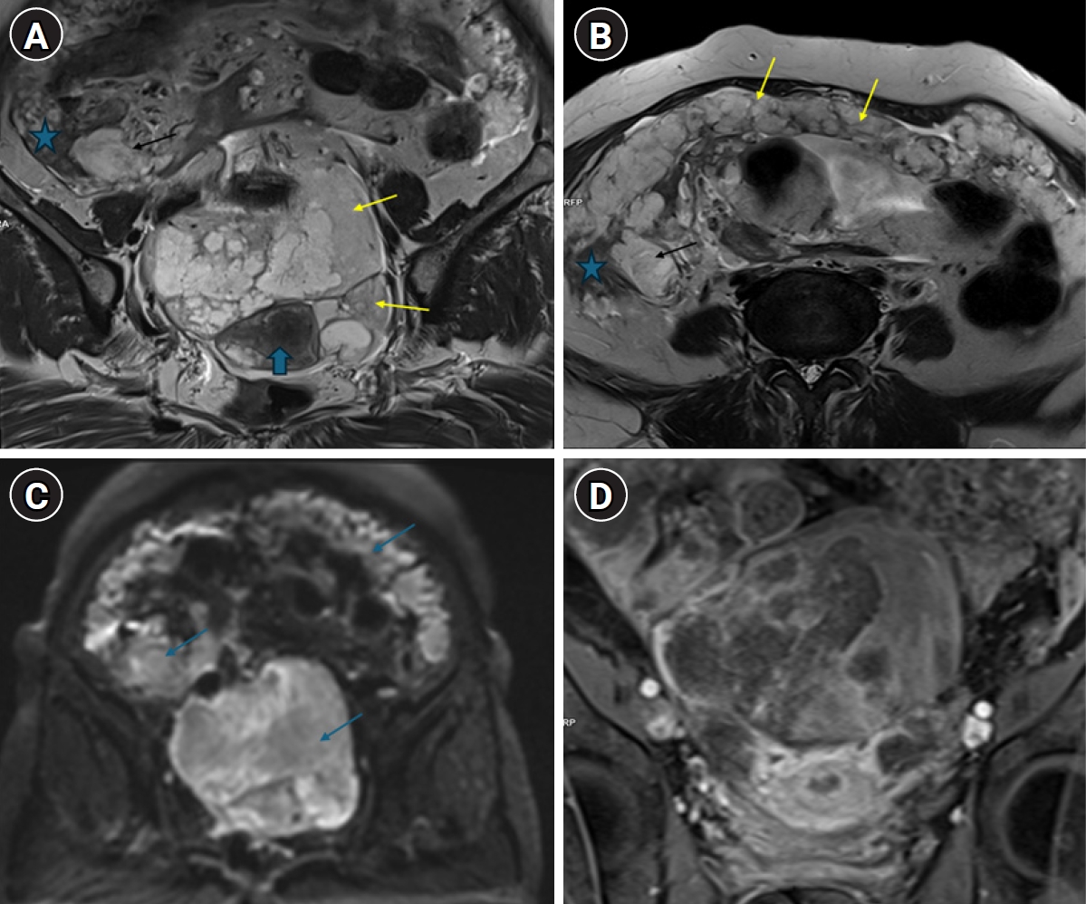
Magnetic resonance imaging (MRI) of the abdomen and pelvis revealed a uterus measuring 80×26×40 mm, with a 23×24-mm intramural fibroid in the fundal region and an ET of 6.3 mm. MRI characterized the lesion as a large, irregular, lobulated, solid-cystic mass measuring 11.5×10×8.9 cm. The mass was T1 hypointense and T2 hyperintense, with diffusion restriction, an apparent diffusion coefficient of 1.2×10
⁻³ mm
2/sec, and heterogeneous contrast enhancement. Neither ovary was visualized separately. Infiltration of the posterior uterine wall, omental caking, and serosal deposits over the cecum and appendix were noted. No significant pelvic lymphadenopathy was seen (
Fig. 1A–
D).
Routine laboratory investigations, including a complete blood count, liver and renal function tests, coagulation profile, blood glucose testing, and viral serology, were within normal limits. Tumor markers showed elevated cancer antigen 125 (CA-125; 213 U/mL) and markedly elevated carcinoembryonic antigen (CEA; 144 ng/mL), whereas carbohydrate antigen 19-9 (CA 19-9) was normal (0.6 U/mL). The Pap smear was negative for intraepithelial lesions.
Management
In view of the findings suggestive of presumed FIGO stage IIIC ovarian malignancy, the patient was scheduled for endometrial biopsy and USG-guided biopsy of the abdominal lesion, followed by 3 cycles of neoadjuvant chemotherapy (NACT) and interval cytoreductive surgery. Histopathological examination of the endometrial biopsy showed atrophic endometrium, whereas biopsy of the abdominal lesion revealed mucin-secreting adenocarcinoma with signet-ring cells. To evaluate a possible GI primary tumor and rule out secondary ovarian involvement, upper GI endoscopy and colonoscopy were performed. Upper GI endoscopy demonstrated grade A esophagitis with gastric erythema, whereas colonoscopy was unremarkable, with no significant abnormalities detected. Immunohistochemical (IHC) analysis was not performed on the preoperative abdominal lesion biopsy specimen because the biopsy material was limited and the initial clinical-radiological impression favored advanced ovarian malignancy.
Moreover, the patient had large bilateral adnexal masses, elevated CA-125 levels, ascites, omental caking, and peritoneal carcinomatosis, all of which strongly suggested advanced epithelial ovarian cancer. Because endoscopic evaluation did not identify a definite GI primary lesion, the patient was started on NACT for presumed primary ovarian malignancy with paclitaxel 250 mg and carboplatin 400 mg every 21 days for 3 cycles. After the second chemotherapy cycle, she developed deep vein thrombosis (DVT) of the right lower limb. Rivaroxaban was initiated at 15 mg twice daily for 21 days, followed by 20 mg once daily. After stabilization, the third chemotherapy cycle was administered as planned.
After completion of 3 cycles of NACT, the patient underwent reassessment. Routine laboratory investigations, including a complete blood count, liver and renal function tests, coagulation profile, and 2-dimensional echocardiography, were within acceptable limits. Venous Doppler examination of both lower limbs showed no residual thrombosis.
Tumor marker evaluation showed an increase in CA-125 to 404 U/mL, whereas CEA decreased to 55.35 ng/mL. Repeat pelvic USG demonstrated a solid, irregularly shaped, heterogeneously echogenic lesion measuring 13.9×9.7×13 cm in the suprapubic region. Nodular hypoechoic plaque-like thickening was noted along the anterior abdominal wall and periphery, suggestive of persistent omental caking. The uterus was visualized separately from the lesion, with an ET of 4 mm.
Table 1 summarizes the clinical, radiological, and biochemical parameters evaluated before and after NACT.
Because no significant radiological response was observed and bulky disease persisted after NACT, interval cytoreductive surgery was planned.
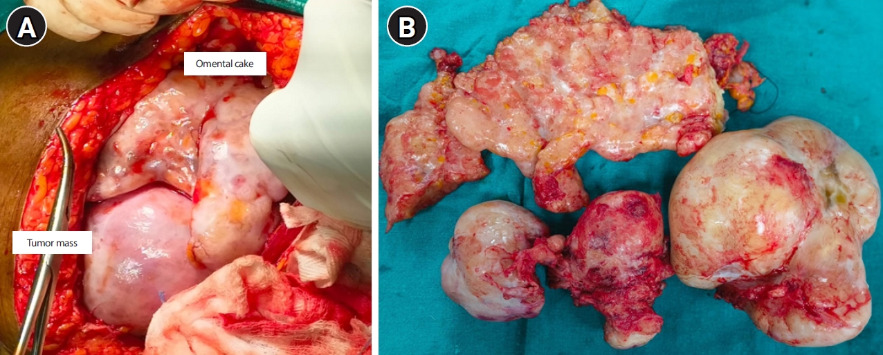
Intraoperative assessment revealed multiple dense adhesions between the bowel loops and the pelvic mass. Mild-to-moderate ascites of approximately 150 mL was present, and approximately 10 mL of peritoneal fluid was aspirated and sent for cytological analysis. A large omental cake measuring approximately 20×18 cm, densely studded with tumor deposits, was adherent to the ovarian mass. A 15×10-cm solid mass was noted arising from the right ovary; it was densely adherent to the posterior uterine surface and adjacent bowel loops, with the right fallopian tube stretched over the mass. A separate 5×6-cm solid mass was identified arising from the left ovary and was densely adherent to the posterior uterine surface, bowel, and left lateral pelvic wall. The uterus appeared bulky, corresponding to approximately 8–10 weeks’ gestational size, and contained a 4×3-cm fundal fibroid (
Fig. 2A,
B). Both round ligaments were thickened, and multiple metastatic deposits were observed over the parietal peritoneum, bowel serosa, appendix, pouch of Douglas, and undersurface of the liver and diaphragm.
Total abdominal hysterectomy with bilateral salpingo-oophorectomy, appendectomy, and infracolic omentectomy was performed. Maximal cytoreduction was performed by removing most visible tumor deposits, except for lesions that densely infiltrated the bowel and liver parenchyma.
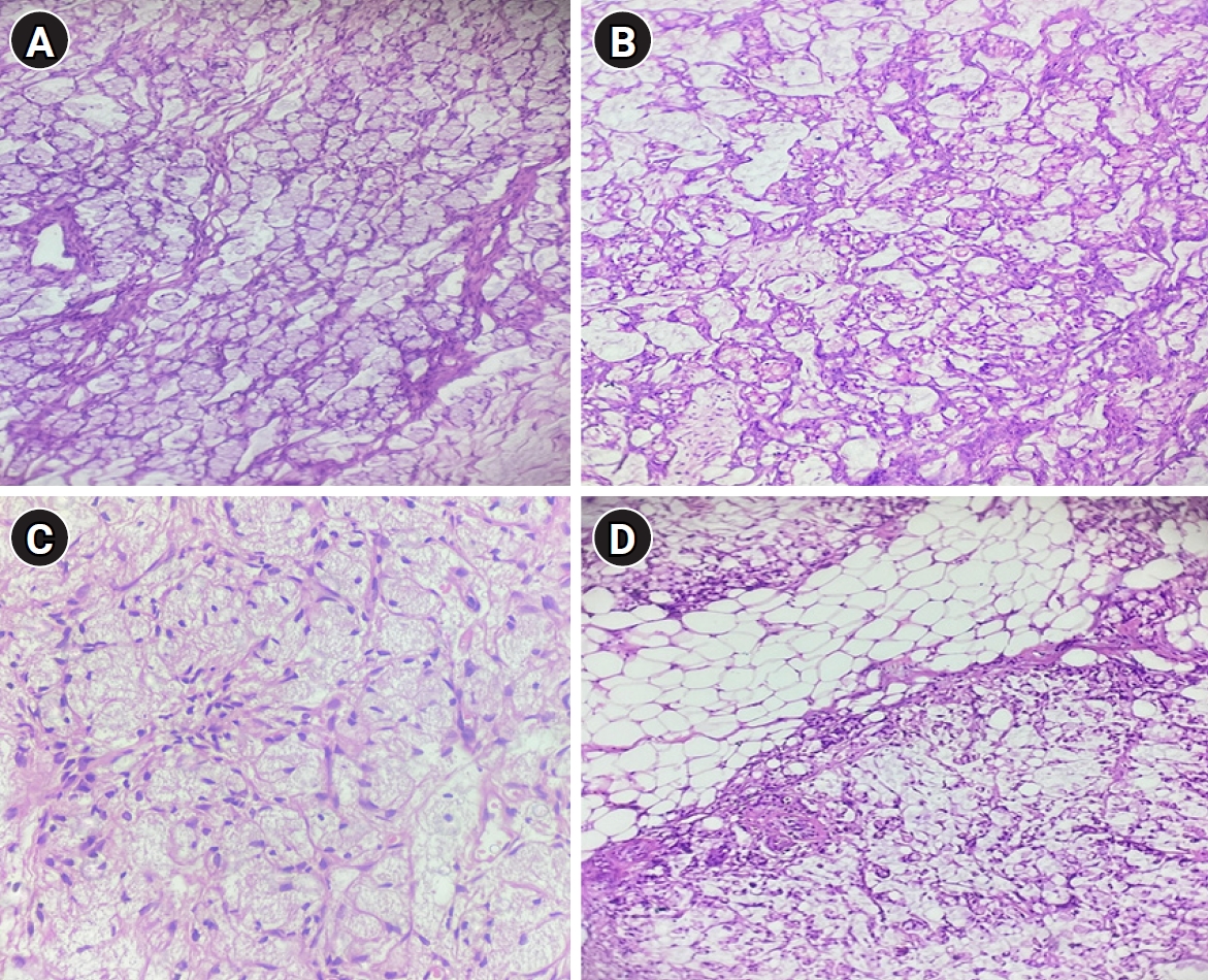
Gross examination revealed an irregular, globular mass involving the right ovary and measuring 15×14×8 cm, with a yellow-to-mucoid cut surface and focal calcification. The left ovarian mass measured 9×7×4.5 cm and had a similar yellow-mucoid solid appearance, with focal hemorrhagic and calcific areas. Microscopic evaluation supported mucinous adenocarcinoma involving both ovaries, with intact capsules.
The right fallopian tube measured 6×0.5 cm and showed serosal deposits without invasion of the tubal wall. The uterus measured 10.5×9×4 cm and had multiple serosal deposits; on cut section, the endometrium measured 2 mm in thickness, and the myometrium measured 2 cm in thickness. The external cervical surface also showed tumor deposits, although the cervical cut section was unremarkable. The left fallopian tube measured 7×0.5 cm and similarly showed serosal deposits without invasion of the tubal wall. A large omental cake measuring 24×14×3 cm was extensively infiltrated by tumor deposits and appeared grayish-yellow. The appendix showed tumor involvement with histological features similar to those of the ovarian masses (
Fig. 3A–
D). In addition, the parietal peritoneum, bilateral parametrium, and all excised deposits showed involvement by mucinous adenocarcinoma. Cytological examination of peritoneal fluid was positive for malignant cells.
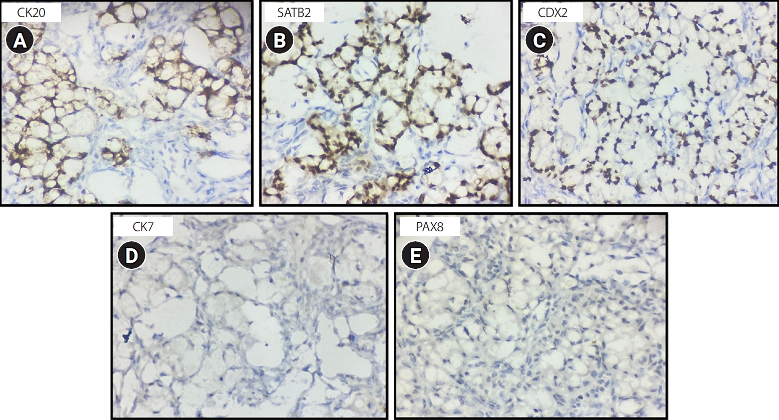
On the basis of these findings, the initial histopathological diagnosis was mucinous adenocarcinoma, grade 2, International Federation of Gynecology and Obstetrics (FIGO) stage IIIC, involving both ovaries. To determine whether the tumor represented a primary ovarian malignancy or secondary ovarian involvement, IHC was performed. Tumor cells showed strong positivity for cytokeratin 20 (CK20), special AT-rich sequence-binding protein 2 (SATB2), and caudal-type homeobox 2 (CDX2), and were negative for cytokeratin 7 (CK7) and paired-box gene 8 (PAX8) (
Fig. 4A–
E). Together with the clinical and radiological findings and the absence of colorectal pathology on colonoscopy, these findings supported a diagnosis of metastatic mucinous adenocarcinoma involving both ovaries, most likely of appendiceal origin.
The patient was discharged in stable condition after suture removal and was subsequently started on 5-fluorouracil (5-FU), leucovorin, and oxaliplatin (FOLFOX) chemotherapy every 14 days. The regimen consisted of oxaliplatin 120 mg intravenously on day 1, leucovorin 550 mg intravenously on day 1, a 5-FU bolus of 550 mg intravenously on day 1, followed by a 5-FU infusion of 3,200 mg over 46 hours. A total of 12 cycles over 6 months was planned, with ongoing clinical and laboratory monitoring to assess tolerance and response. The patient is currently undergoing regular follow-up and has completed 9 cycles of FOLFOX chemotherapy. She is tolerating chemotherapy well, without major adverse effects, and has had significant improvement in abdominal symptoms and ascites. Positron emission tomography/computed tomography is planned after completion of the final chemotherapy cycle to evaluate treatment response and current disease status.
Ethics statement
According to institutional policy, ethical approval is not required for single case reports. Written informed consent was obtained for publication of this case and the accompanying clinical data and images.
Discussion
Metastatic tumors involving the ovary account for approximately 10%–25% of all ovarian malignancies [
5]. They arise from a wide range of primary sites, most commonly the breast, colorectum, stomach, and endometrium, with less frequent origins including the small intestine, appendix, pancreas, biliary tract, and lung [
6].
Appendiceal cancer is rare, with an incidence of 1–2 per million, and encompasses a heterogeneous spectrum ranging from low-grade appendiceal mucinous neoplasms to high-grade adenocarcinomas [
7,
8]. According to the Surveillance, Epidemiology, and End Results (SEER) database, epithelial adenocarcinoma is the most frequent broad histologic category, with mucinous adenocarcinoma representing the predominant epithelial subtype (37%) [
5,
9]. In contrast, when all appendiceal neoplasms are considered irrespective of histology, carcinoid (neuroendocrine) tumors are the most common, accounting for approximately 66% of cases, followed by cystadenocarcinomas (20%) and adenocarcinomas (10%) [
10].
The most frequent pattern of spread involves the peritoneal cavity, resulting in extensive mucinous dissemination and peritoneal metastases [
11]. Clinically, appendiceal malignancies commonly present as acute appendicitis, although incidental detection during surgery, pelvic abscess, GI symptoms, and bowel obstruction have also been described [
4,
5]. In the present case, the patient had a prior history of acute appendicitis, for which appendiceal biopsy was performed; however, histopathological examination revealed only nonspecific inflammatory changes, with no evidence of malignancy. The original histopathology slides or blocks were unavailable for re-review; therefore, the possibility that an occult appendiceal malignancy was missed at that time cannot be completely excluded. This limitation illustrates an important diagnostic challenge associated with appendiceal neoplasms, particularly when they initially mimic benign inflammatory disease.
Bilateral ovarian involvement in appendiceal mucinous adenocarcinoma is well recognized and may closely resemble primary ovarian malignancy, creating substantial diagnostic difficulty [
12]. Differentiating primary appendiceal mucinous neoplasms from primary ovarian mucinous tumors is particularly challenging in advanced or clinically complex cases. Preoperative evaluation is frequently inconclusive because GI endoscopy has limited sensitivity for appendiceal lesions [
9,
12], and currently available tumor biomarkers, including CEA, CA 19-9, and CA-125, lack specificity because they may be elevated in both GI and ovarian malignancies [
4]. Moreover, metastatic appendiceal mucinous adenocarcinomas can mimic primary ovarian mucinous carcinomas in clinical presentation, imaging characteristics, and gross morphology, often requiring definitive histopathological and IHC evaluation for accurate diagnosis [
4,
12]. Notably, emerging evidence indicates that true primary mucinous ovarian carcinomas constitute only a small proportion, approximately 3%, of ovarian malignancies, supporting the view that many mucinous ovarian tumors are metastatic in origin [
13].
Clinically, patients often present with vague, nonspecific abdominal symptoms and imaging findings suggestive of advanced ovarian cancer. These features further complicate preoperative differentiation because appendiceal primaries are rare and radiological findings are often nonspecific [
4]. The absence of GI symptoms, together with abdominal distension and elevated CA-125 levels, may further favor an erroneous diagnosis of primary ovarian cancer [
10]. In many cases, ovarian metastases become clinically apparent before the primary tumor is identified, which may delay determination of the tumor origin and selection of the most appropriate management strategy [
5]. Typical presenting complaints include abdominal pain (42%), postmenopausal bleeding (18%), and abdominal bloating (15%), whereas only a minority of patients have symptoms directly attributable to the primary lesion [
5,
14]. Although bilateral ovarian masses may raise suspicion for metastatic disease, similar findings may also occur in some primary ovarian tumors, including undifferentiated and serous carcinomas, underscoring the need for comprehensive pathological evaluation [
5,
15,
16].
In the present case, the patient presented with an abdominal mass accompanied by vague symptoms, including abdominal pain, distension, and loss of appetite, without altered bowel habits or rectal bleeding. The initial appendiceal biopsy showed only nonspecific inflammation, and both upper GI endoscopy and colonoscopy were unremarkable, further complicating preoperative diagnosis. Consequently, NACT followed by interval debulking surgery was considered, as is commonly recommended for advanced ovarian tumors [
17]. However, the patient did not have a satisfactory response to neoadjuvant therapy, prompting further evaluation and revision of the diagnosis.
The presence of signet-ring cells in appendiceal tumors, even in small proportions, has been consistently associated with aggressive tumor biology and poorer survival outcomes [
18,
19]. For appropriately selected patients with peritoneal dissemination, cytoreductive surgery combined with hyperthermic intraperitoneal chemotherapy (CRS-HIPEC) remains an important treatment strategy; outcomes are influenced by disease burden, completeness of cytoreduction, histological subtype, and tumor biology [
7]. In the present case, surgery was performed for presumed advanced ovarian malignancy on the basis of the clinical, radiological, and intraoperative findings. Because the final histopathological diagnosis of appendiceal mucinous adenocarcinoma was not known at the time of surgery, CRS-HIPEC was not considered. After definitive histopathological and IHC confirmation, the patient was managed with systemic chemotherapy. Current systemic treatment for advanced appendiceal adenocarcinoma is largely extrapolated from colorectal cancer protocols and commonly includes regimens such as FOLFOX or FOLFIRI, with or without targeted agents such as bevacizumab or cetuximab [
20].
In conclusion, this case highlights the diagnostic challenge posed by appendiceal mucinous adenocarcinoma presenting as bilateral ovarian masses and clinically mimicking advanced primary ovarian malignancy. The absence of GI symptoms, nonspecific imaging findings, and elevated CA-125 levels initially supported a gynecologic primary tumor, leading to NACT; however, the lack of therapeutic response prompted further evaluation. A definitive diagnosis was achieved only after histopathological examination and IHC supported a diagnosis of metastatic mucinous adenocarcinoma involving both ovaries, most consistent with an appendiceal primary. This case underscores the importance of considering secondary ovarian tumors in patients with bilateral mucinous ovarian masses, particularly when tumor markers such as CEA are markedly elevated or the clinical response is atypical.
-
Authors’ contribution
Conceptualization: NK. Literature search: NK. Data collection: NK, GR, AS, MM, NB, PTR. Formal analysis: NK, GR, AS, MM, NB, PTR. Writing–original draft: NK. Writing–review and editing: NK, GR, AS, MM, NB, PTR. Final approval of the manuscript: all authors.
-
Conflict of interest
No potential conflict of interest relevant to this article was reported.
-
Funding
None.
-
Data availability
Not applicable.
-
Acknowledgments
None.
-
Supplementary materials
None.
Fig. 1.Magnetic resonance imaging findings. (A) Coronal T2-weighted image showing bilateral T2-hyperintense ovarian mass lesions (yellow arrows). The mass (black arrow) infiltrates the cecum (blue asterisk). The appendix is not separately visualized. The blue block arrow indicates the uterus. (B) Axial T2-weighted image showing a tumor deposit (black arrow) infiltrating the cecum (blue asterisk), with omental deposits resulting in caking (yellow arrows). (C) Axial diffusion-weighted image at a high b-value of 1,000 s/mm² showing restricted diffusion in the omentum, ovarian mass lesions, and cecal deposit. (D) Coronal contrast-enhanced T1-weighted fat-saturated image showing heterogeneous enhancement of the ovarian mass.
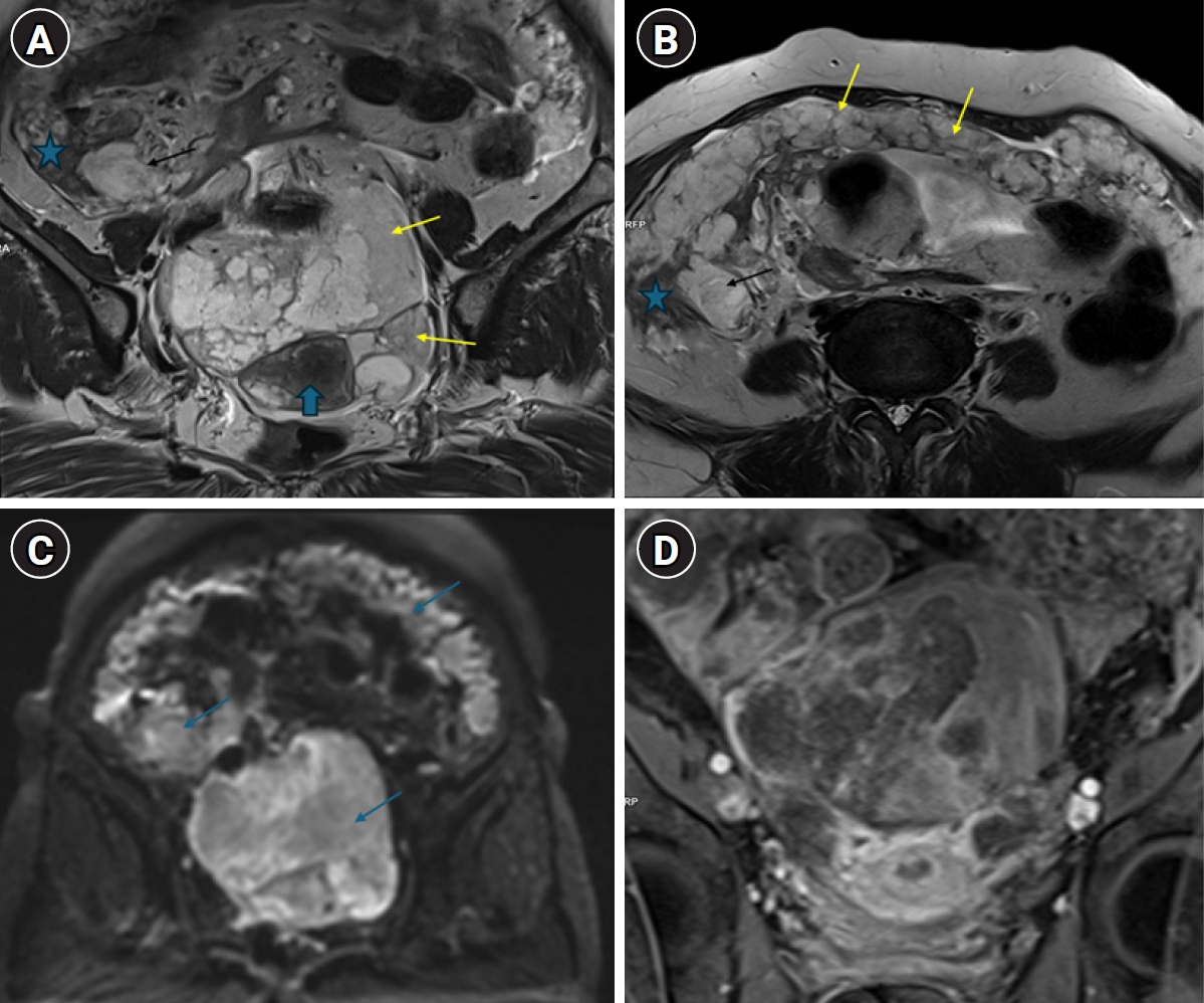
Fig. 2.Intraoperative and specimen findings. (A) Intraoperative finding showing a large omental cake and tumor mass. (B) Surgical specimen showing the uterus and cervix, bilateral ovarian masses, omental cake, and appendix.
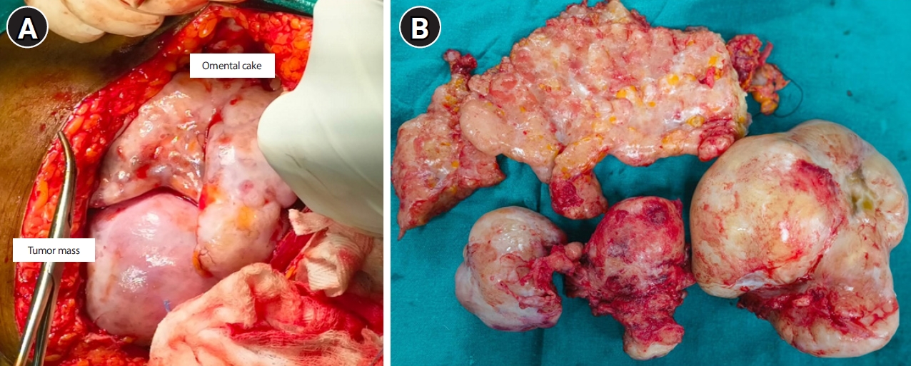
Fig. 3.Histopathological findings. (A) Mucinous adenocarcinoma involving the right ovary demonstrating intracellular mucin, marked nuclear atypia, prominent nucleoli, and abnormal mitotic activity in tumor cells (hematoxylin and eosin [H&E], ×10). (B) Section of mucinous adenocarcinoma involving the left ovary showing complex glandular architecture with stratified atypical mucin-secreting epithelial cells, nuclear pleomorphism, and stromal invasion (H&E, ×10). (C) Section of the appendix showing tumor cells with abundant intracellular and extracellular mucin and floating tumor cells with signet-ring cell morphology (H&E, ×10). (D) Section showing fibromuscular and collagenous omental tissue infiltrated by malignant tumor cells of mucinous adenocarcinoma (H&E, ×10).
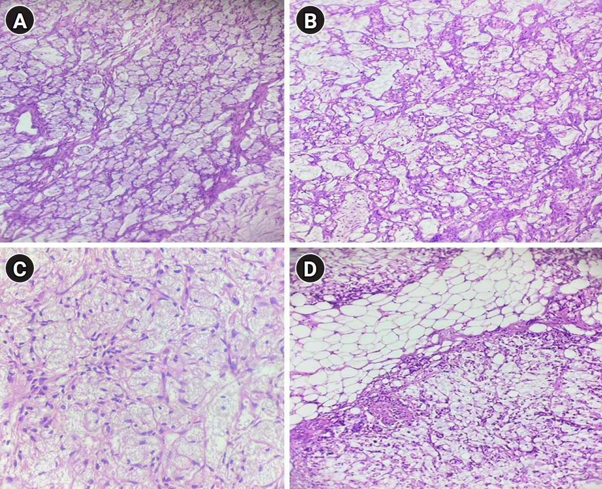
Fig. 4.Immunohistochemical analysis. (A) Immunohistochemical (IHC) staining showing diffuse cytoplasmic positivity for cytokeratin 20 (CK20). (B) IHC staining showing strong nuclear positivity for special AT-rich sequence-binding protein 2 (SATB2) in tumor cells. (C) IHC staining showing strong nuclear positivity for caudal-type homeobox 2 (CDX2) in tumor cells. (D) IHC staining showing absence of cytokeratin 7 (CK7) expression in tumor cells. (E) IHC staining showing absence of paired-box gene 8 (PAX8) expression in tumor cells.
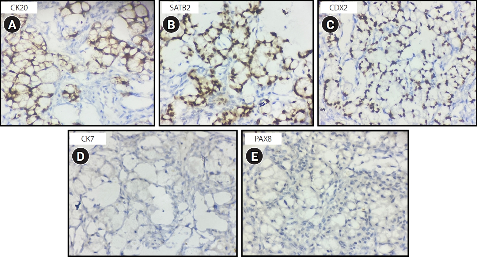
Table 1.Comparison of investigational findings before and after neoadjuvant chemotherapy
|
Investigation |
Pre-NACT findings |
Post-NACT findings/reassessment |
Inference/response |
|
Pelvic USG |
Large heterogeneously hypoechoic solid pelvic mass measuring 15.9×9.7×11 cm, with internal vascularity; ovaries not visualized separately; mild ascites |
Solid, irregular, heterogeneously echogenic lesion measuring 13.9×9.7×13 cm; nodular plaque-like thickening suggestive of persistent omental caking; uterus visualized separately; ET 4 mm |
No significant size reduction; persistent bulky disease |
|
CECT of the abdomen and pelvis |
Lobulated heterogeneously hypodense pelvic mass measuring 10.6×9.9×8.9 cm, with peripheral enhancement and necrotic areas; moderate ascites; omental caking; loss of fat planes; ovaries not visualized separately |
Not repeated |
Baseline advanced disease |
|
MRI of the abdomen and pelvis |
Large, irregular, lobulated, solid-cystic mass measuring 11.5×10×8.9 cm; T1 hypointense and T2 hyperintense, with diffusion restriction; posterior uterine wall infiltration; serosal deposits over the cecum and appendix; omental caking |
Not repeated |
Extensive baseline disease burden |
|
CA-125 |
213 U/mL |
Increased to 404 U/mL |
Biochemical progression |
|
CEA |
144 ng/mL |
Decreased to 55.35 ng/mL |
Partial biochemical response; supports a GI primary |
|
CA 19-9 |
Normal, 0.6 U/mL |
Not repeated |
- |
|
Venous Doppler |
Not applicable |
No residual thrombosis after DVT treatment |
Stabilized complication |
References
- 1. Dundr P, Singh N, Nožičková B, Němejcová K, Bártů M, Stružinská I. Primary mucinous ovarian tumors vs. ovarian metastases from gastrointestinal tract, pancreas and biliary tree: a review of current problematics. Diagn Pathol 2021;16:20. https://doi.org/10.1186/s13000-021-01079-2
- 2. Kawecka W, Pasnik I, Adamiak-Godlewska A, Semczuk M, Tyczynska M, Semczuk A. How to differentiate primary mucinous ovarian tumors from ovarian metastases originating from primary appendiceal mucinous neoplasms: a review. Pathol Oncol Res 2025;31:1612066. https://doi.org/10.3389/pore.2025.1612066
- 3. Yu Y, Wang T, Yuan Z, Lin W, Yang J, Cao D. Misdiagnosed appendiceal mucinous neoplasms and primary ovarian mucinous tumors present with different pre- and intraoperative characteristics. Front Oncol 2022;12:966844. https://doi.org/10.3389/fonc.2022.966844
- 4. Perivoliotis K, Christodoulidis G, Samara AA, Sgantzou IK, Floros T, Volakakis G, Karasavvidou F, Tepetes K. Low-grade appendiceal mucinous neoplasm (LAMN) primarily diagnosed as an ovarian mucinous tumor. Case Rep Surg 2021;2021:5523736. https://doi.org/10.1155/2021/5523736
- 5. Lin CW, Peng SL, Wang SH, Wu PY. A case report of appendiceal adenocarcinoma without gastrointestinal evidence mimicking primary ovarian cancer. J Int Med Res 2022;50:3000605221088559. https://doi.org/10.1177/03000605221088559
- 6. Zhang JJ, Cao DY, Yang JX, Shen K. Ovarian metastasis from nongynecologic primary sites: a retrospective analysis of 177 cases and 13-year experience. J Ovarian Res 2020;13:128. https://doi.org/10.1186/s13048-020-00714-8
- 7. Strach MC, Sutherland S, Horvath LG, Mahon K. The role of chemotherapy in the treatment of advanced appendiceal cancers: summary of the literature and future directions. Ther Adv Med Oncol 2022;14:17588359221112478. https://doi.org/10.1177/17588359221112478
- 8. Dsouza R, Osueni A, Menon G. Appendiceal tumors [Internet]. StatPearls Publishing; 2025 [cited 2026 Feb 6]. Available from: https://www.ncbi.nlm.nih.gov/books/NBK555943/
- 9. Leonards LM, Pahwa A, Patel MK, Petersen J, Nguyen MJ, Jude CM. Neoplasms of the appendix: pictorial review with clinical and pathologic correlation. Radiographics 2017;37:1059-1083. https://doi.org/10.1148/rg.2017160150
- 10. Boyajian H, Majeski V, Flores A, Sturtz D, Baidoun F, Dughayli M. Clinicopathological and Perioperative Outcome of Appendiceal Tumors: Case Review of 31 Patients. Spartan Med Res J 2020;5:13487. https://doi.org/10.51894/001c.13487
- 11. Carr NJ, Bibeau F, Bradley RF, Dartigues P, Feakins RM, Geisinger KR, Gui X, Isaac S, Milione M, Misdraji J, Pai RK, Rodriguez-Justo M, Sobin LH, van Velthuysen MF, Yantiss RK. The histopathological classification, diagnosis and differential diagnosis of mucinous appendiceal neoplasms, appendiceal adenocarcinomas and pseudomyxoma peritonei. Histopathology 2017;71:847-858. https://doi.org/10.1111/his.13324
- 12. Zhang W, Tan C, Xu M, Wu X. Appendiceal mucinous neoplasm mimics ovarian tumors: challenges for preoperative and intraoperative diagnosis and clinical implication. Eur J Surg Oncol 2019;45:2120-2125. https://doi.org/10.1016/j.ejso.2019.08.004
- 13. Yemelyanova AV, Vang R, Judson K, Wu LS, Ronnett BM. Distinction of primary and metastatic mucinous tumors involving the ovary: analysis of size and laterality data by primary site with reevaluation of an algorithm for tumor classification. Am J Surg Pathol 2008;32:128-138. https://doi.org/10.1097/PAS.0b013e3180690d2d
- 14. Lobo J, Machado B, Vieira R, Bartosch C. The challenge of diagnosing a malignancy metastatic to the ovary: clinicopathological characteristics vary and morphology can be different from that of the corresponding primary tumor. Virchows Arch 2017;470:69-80. https://doi.org/10.1007/s00428-016-2029-3
- 15. Foti PV, Attinà G, Spadola S, Caltabiano R, Farina R, Palmucci S, Zarbo G, Zarbo R, D'Arrigo M, Milone P, Ettorre GC. MR imaging of ovarian masses: classification and differential diagnosis. Insights Imaging 2016;7:21-41. https://doi.org/10.1007/s13244-015-0455-4
- 16. Willmott F, Allouni KA, Rockall A. Radiological manifestations of metastasis to the ovary. J Clin Pathol 2012;65:585-590. https://doi.org/10.1136/jclinpath-2011-200390
- 17. Vergote I, Coens C, Nankivell M, Kristensen GB, Parmar MKB, Ehlen T, Jayson GC, Johnson N, Swart AM, Verheijen R, McCluggage WG, Perren T, Panici PB, Kenter G, Casado A, Mendiola C, Stuart G, Reed NS, Kehoe S; EORTC; MRC CHORUS study investigators. Neoadjuvant chemotherapy versus debulking surgery in advanced tubo-ovarian cancers: pooled analysis of individual patient data from the EORTC 55971 and CHORUS trials. Lancet Oncol 2018;19:1680-1687. https://doi.org/10.1016/S1470-2045(18)30566-7
- 18. Levinsky NC, Morris MC, Wima K, Sussman JJ, Ahmad SA, Cloyd JM, Kimbrough C, Fournier K, Lee A, Dineen S, Dessureault S, Veerapong J, Baumgartner JM, Clarke C, Zaidi MY, Staley CA, Maithel SK, Leiting J, Grotz T, Lambert L, Hendrix RJ, Ronnekleiv-Kelly S, Pokrzywa C, Raoof M, Eng OS, Johnston FM, Greer J, Patel SH. Should we be doing cytoreductive surgery with HIPEC for signet ring cell appendiceal adenocarcinoma?: a study from the US HIPEC Collaborative. J Gastrointest Surg 2020;24:155-164. https://doi.org/10.1007/s11605-019-04336-4
- 19. Solomon D, DeNicola N, Feingold D, Liu PH, Aycart S, Golas BJ, Sarpel U, Labow DM, Magge DR. Signet ring cell features with peritoneal carcinomatosis in patients undergoing cytoreductive surgery and hyperthermic intraperitoneal chemotherapy are associated with poor overall survival. J Surg Oncol 2019;119:758-765. https://doi.org/10.1002/jso.25379
- 20. Hoehn RS, Rieser CJ, Choudry MH, Melnitchouk N, Hechtman J, Bahary N. Current management of appendiceal neoplasms. Am Soc Clin Oncol Educ Book 2021;41:1-15. https://doi.org/10.1200/EDBK_321009


 , Gunvanti Rathod2