A 77-year-old nursing home resident with hypertension and chronic obstructive pulmonary disease presented to the emergency department with fever and dyspnea of 2 days’ duration. No choking episode had been witnessed, and SARS-CoV-2 (severe acute respiratory syndrome coronavirus 2) testing was positive. On examination, he was tachypneic, with a respiratory rate of 32 breaths/min, an oxygen saturation of 88% on room air, and markedly diminished breath sounds over the left hemithorax.
Laboratory tests showed a white blood cell count of 6,900/μL with 15% band forms, a C-reactive protein level of 163 mg/L, and a procalcitonin level of 0.05 ng/mL. Arterial blood gas analysis while the patient was receiving 10 L/min of oxygen showed alkalemia, with a pH of 7.55, PaCO
2 of 43 mm Hg, and HCO
3⁻ of 36.5 mmol/L, as well as hypoxemia, with a PaO
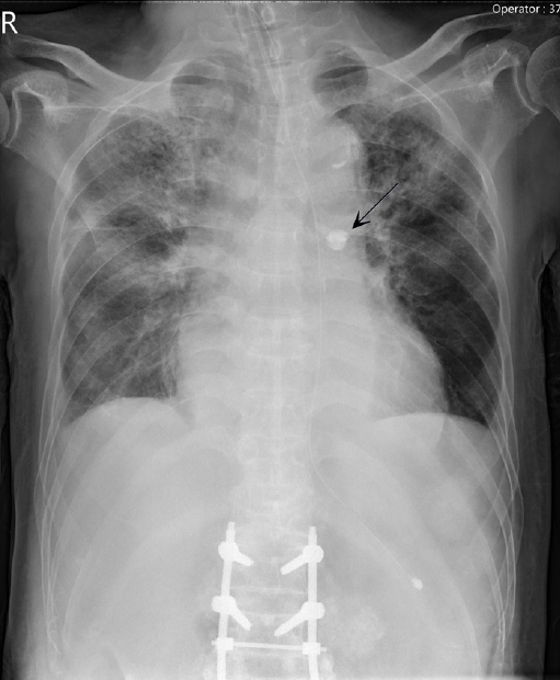
2 of 71 mm Hg. A portable chest radiograph showed bilateral infiltrates and pleural effusions; it also showed hyperinflation of the left lung (
Fig. 1). This unilateral hyperinflation was later attributed to a check-valve mechanism caused by the aspirated tooth in the left main bronchus: airflow entered the distal airways through the partially obstructed bronchus during inspiration, but expiratory airflow was impeded, causing air trapping. This radiographic pattern is a characteristic clue to foreign body aspiration. Thoracentesis yielded a sterile, lymphocyte-predominant transudative effusion, consistent with viral pleuritis and volume overload rather than empyema.
Despite treatment for coronavirus disease 2019 (COVID-19), severe hypoxemia and left-sided hypoventilation persisted. On hospital day 23, re-evaluation of the admission radiograph revealed a subtle radiopaque structure superimposed on the left hilum (
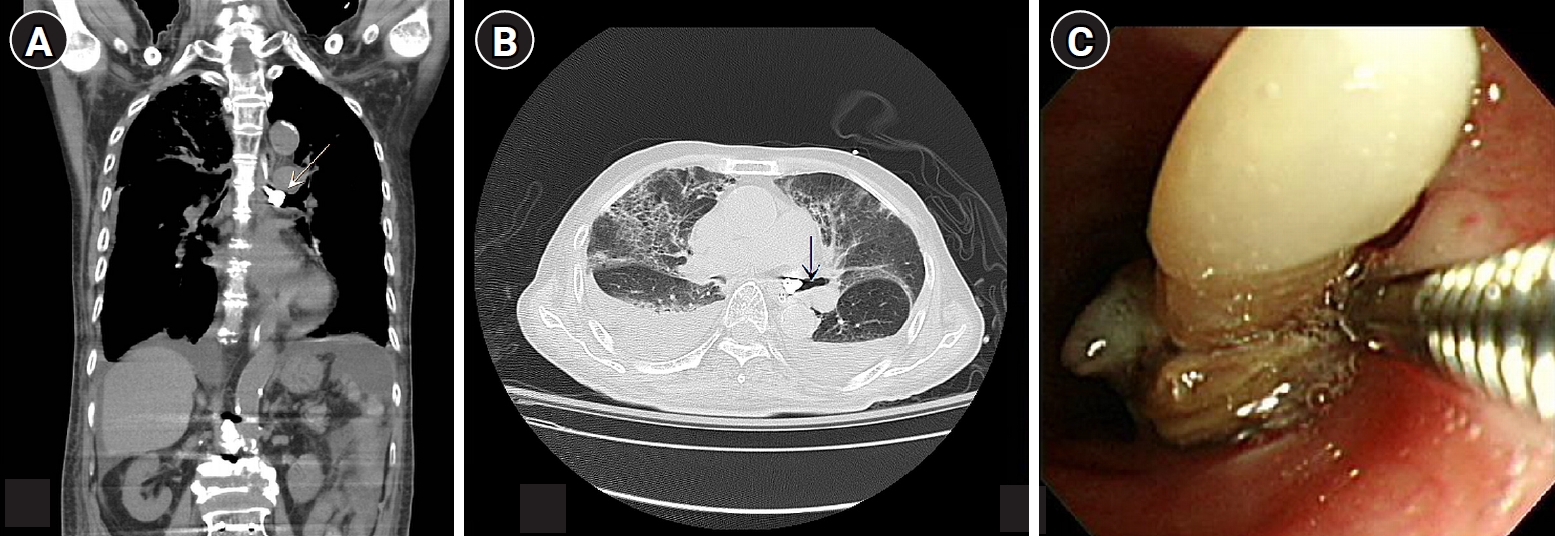
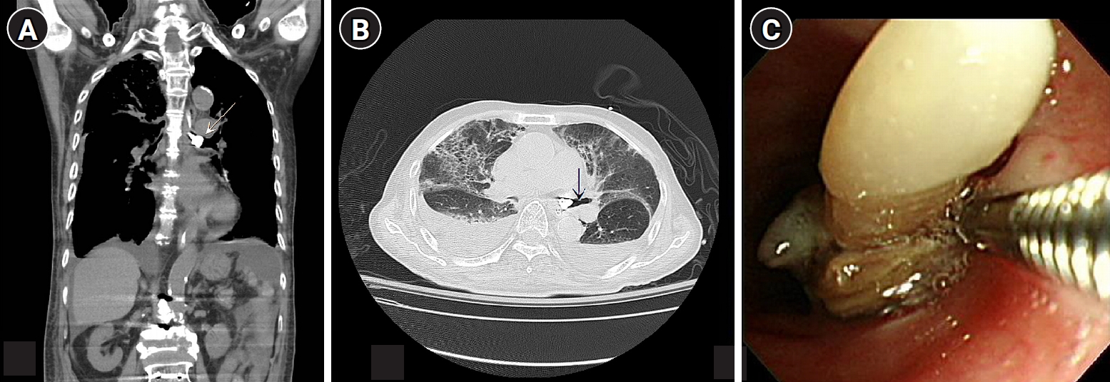
Fig. 1, arrow) that had initially been dismissed as benign calcification. Subsequent chest computed tomography showed an aspirated tooth in the left main bronchus, causing complete obstruction and atelectasis (
Fig. 2A). Lung-window images demonstrated distal air trapping compatible with a check-valve mechanism caused by the aspirated tooth (
Fig. 2B). Flexible bronchoscopy revealed the tooth embedded in granulation tissue (
Fig. 2C). Removal briefly improved oxygenation, but the clinical course was complicated by postobstructive pneumonia, methicillin-resistant
Staphylococcus aureus bacteremia, and multidrug-resistant
Acinetobacter baumannii infection. The patient died on hospital day 106.
The requirement for informed consent was waived by the Institutional Review Board (IRB No. 202501221B0).
Foreign body aspiration in adults is frequently unwitnessed and may be diagnosed without a history of choking, particularly in older adults with dentures or diminished airway reflexes [
1]. In this case, the aspirated tooth mimicked a calcified hilar lesion: projectional superimposition on a portable radiograph made the tooth resemble a calcified hilar lymph node.
The diagnostic delay was likely amplified by anchoring bias. After COVID-19 was confirmed, persistent hypoxemia was attributed to viral pneumonia, and the discordant bedside finding of asymmetric breath sounds was rationalized rather than investigated [
2]. Persistent unilateral hypoventilation that is disproportionate to radiographic findings should prompt urgent evaluation for bronchial obstruction [
3]. When clinical improvement stalls, deliberate repeat review of the initial imaging—with contrast and window settings adjusted to assess the central airways—may reveal a reversible cause before fatal complications develop.
-
Authors’ contribution
Conceptualization: PYC, YJS. Data curation: PYC, CWC. Investigation: PYC, CWC. Methodology: PYC. Validation: CWC. Project administration: YJS. Resources: CWC. Visualization: YJS. Supervision: YJS. Writing–original draft: PYC, CWC. Writing–review & editing: YJS.
-
Conflict of interest
No potential conflict of interest relevant to this article was reported.
-
Funding
None.
-
Data availability
Not applicable.
-
Acknowledgments
None.
-
Supplementary materials
None.
Fig. 1.A portable anteroposterior chest radiograph showing bilateral infiltrates and pleural effusions. A radiopaque foreign body (arrow) is visible over the left hilum, with hyperinflation of the left lung due to distal air trapping.
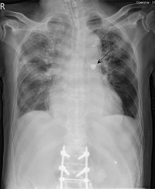
Fig. 2.Coronal chest computed tomography shows a tooth (arrow) causing complete obstruction of the left main bronchus with distal lung collapse (A). Lung-window imaging demonstrates air trapping compatible with a check-valve mechanism caused by the aspirated tooth (arrow) (B). Flexible bronchoscopy shows the tooth embedded in granulation tissue during retrieval (C).
References
- 1. Hewlett JC, Rickman OB, Lentz RJ, Prakash UB, Maldonado F. Foreign body aspiration in adult airways: therapeutic approach. J Thorac Dis 2017;9:3398-3409. https://doi.org/10.21037/jtd.2017.06.137
- 2. Croskerry P. The importance of cognitive errors in diagnosis and strategies to minimize them. Acad Med 2003;78:775-780. https://doi.org/10.1097/00001888-200308000-00003
- 3. Pinto A, Scaglione M, Pinto F, Guidi G, Pepe M, Del Prato B, Grassi R, Romano L. Tracheobronchial aspiration of foreign bodies: current indications for emergency plain chest radiography. Radiol Med 2006;111:497-506. https://doi.org/10.1007/s11547-006-0045-0
Citations
Citations to this article as recorded by